Figure 2. Localization of the mutant EPHA2-Myc proteins in MDCK cells. The ectopically expressed EPHA2-Myc protein (red) in MDCK cells
was detected using anti-Myc primary antibody and Alexa Flour 594-conjugated secondary antibody. Nuclei (blue) were labeled
with DAPI. Cells expressing the wild-type EPHA2-Myc (A) and p.P584L (B), p.A959T (E), or pV972fsX39 (F) mutant EPHA2-Myc proteins show peripheral and cytoplasmic localization of the protein. In cells expressing the p.T940I (C) and p.D942fsXC71 (D) mutant EPHA2-Myc proteins, the protein can be seen predominantly in the perinuclear region (arrow) and some in the cytoplasm.
Mock transfected cells were used as a negative control (G). Representative images from two independent experiments are shown. Scale-bar=20 µm.
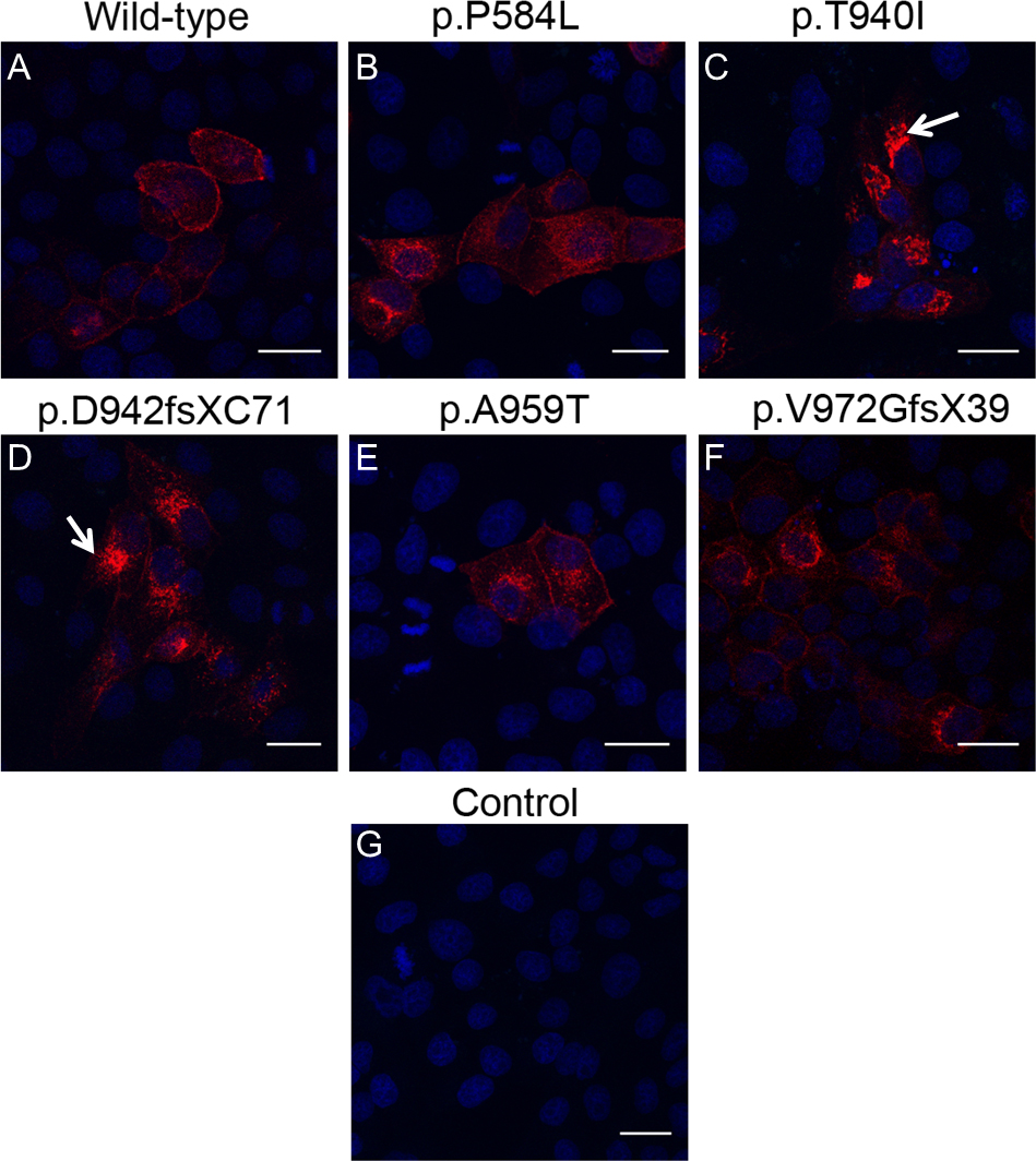
 Figure 2 of
Dave, Mol Vis 2016; 22:18-30.
Figure 2 of
Dave, Mol Vis 2016; 22:18-30.