Figure 3. Colocalization of Krt4 and Krt10. A: Sagittal section of the eye immunostained for either keratin 10 (Krt10) or Krt4 in a P21 mouse. Note the distinct expression
pattern of these two lineage markers; Krt4 was expressed in the meibomian gland (inset) and the conjunctiva, and Krt10 was
restricted to the epidermis. B: Ten-micron horizontal cryosections double stained for Krt4 (red) and Krt10 (green). Inset shows the meibomian gland with
a small colocalization region at P2 and a larger degree of colocalization at P4 in the developing duct. By P11, the areas
of colocalization were reduced with only a small region present within the central duct. C: Fifty-micron sections double stained for Krt4 (red) and Krt10 (green) at P4 and P11 with a three-dimensional composite (bottom
panel). MG = meibomian gland, EP = epidermis; MCJ = mucocutaneous junction; CJ = conjunctiva; asterisk = central duct; arrow
= elongated epidermal invagination.
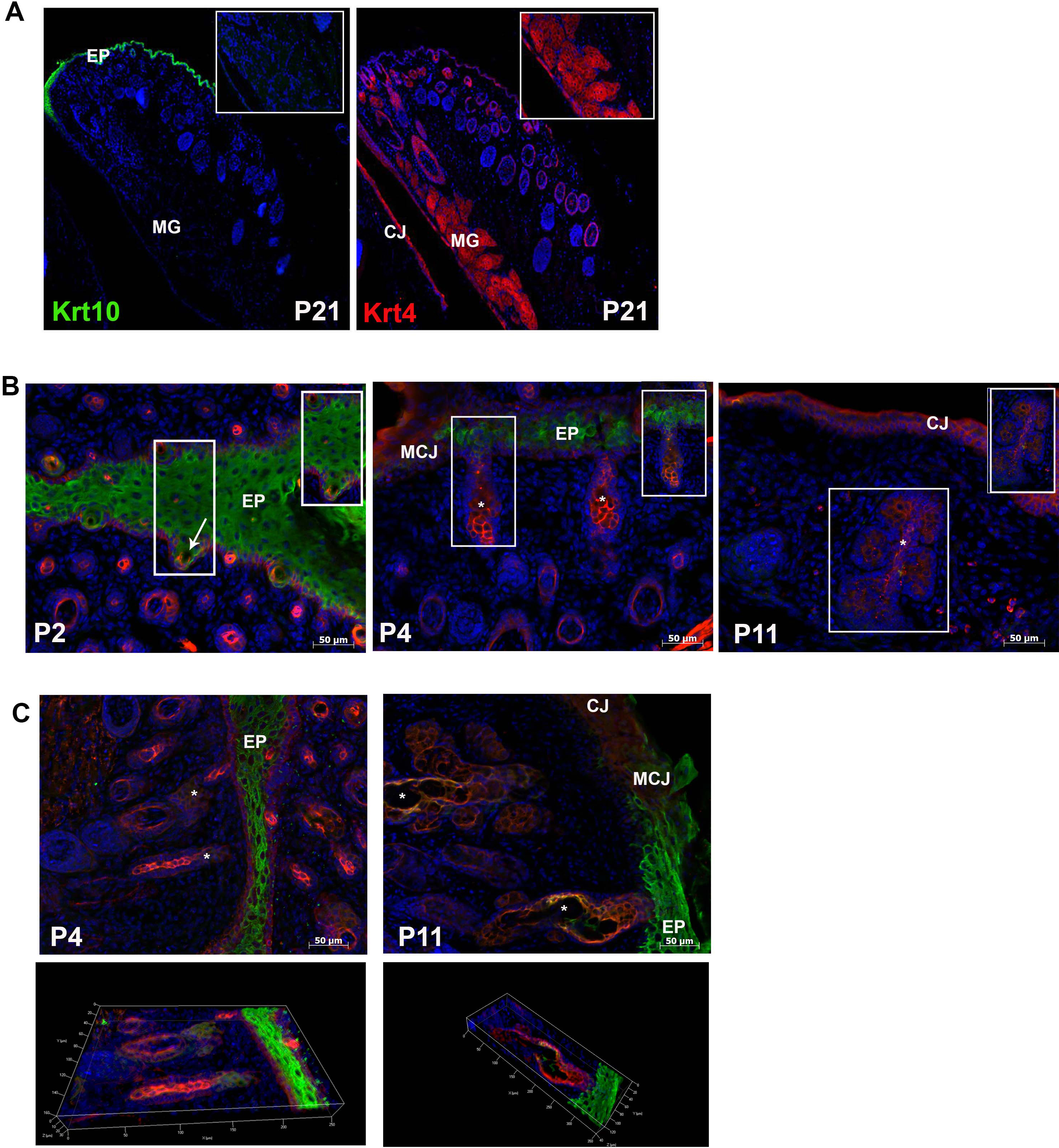
 Figure 3 of
Call, Mol Vis 2016; 22:168-176.
Figure 3 of
Call, Mol Vis 2016; 22:168-176.