Figure 2. SST reduces calpain-2 activation and PTP1B expression and cleavage induced by high glucose and directly activates IGF-IR/Akt
phosphorylation in 661W cells and retinal explants. The 661W cells were cultured under basal conditions (B) or with medium
supplemented with 55 mM glucose (high glucose, HG) during 24 h in the absence or presence of 10−6 M somatostatin (SST). Levels of the calpain-2 proform (80-kDa) and the 58-kDa fragment (A) and the levels of protein tyrosine phosphatase 1B (PTP1B) (B) were analyzed with western blot with β-actin as a loading control. The graphs correspond to the quantification and statistical
analysis of data obtained in three independent experiments performed in duplicate. *p<0.05 versus basal or †p<0.05 versus
HG condition. The 661W cells (C) and the retinal explants (D) were treated with SST (10−6 M) for 24 h or left untreated. At the end of the culture time, the phosphorylation levels of type 1 insulin-like growth factor
receptor (IGF-IR; tyrosine 1165/66) and protein kinase B (Akt; serine 473) were analyzed with western blot. Results were normalized
with respect to the total IGF-IR or Akt levels. The graphs correspond to the quantification and statistical analysis of data
obtained in three independent experiments performed in duplicate (C) or five retinas per condition (D). *p<0.05 versus Basal.
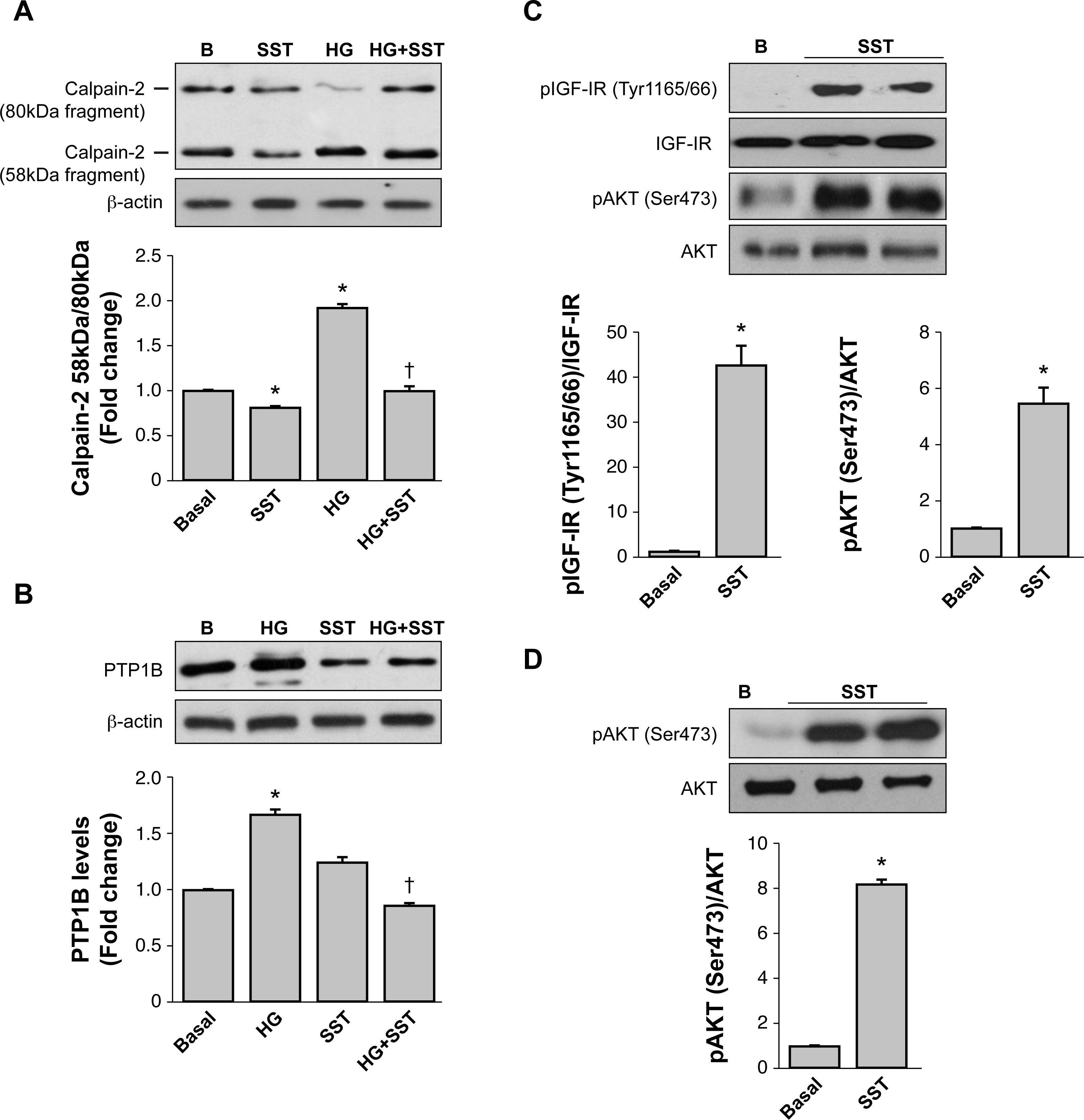
 Figure 2 of
Arroba, Mol Vis 2016; 22:1522-1531.
Figure 2 of
Arroba, Mol Vis 2016; 22:1522-1531.