Figure 4. Nrf2-deficient EAE mice exhibit more severe inflammation of the optic nerve and demyelination. A: Hematoxylin and eosin (H&E)-stained, paraffin-embedded optic nerves from wild-type (WT; blue circles) or knockout (KO; red
squares) experimental autoimmune encephalomyelitis (EAE) mice were scored on a scale of 0–3 for inflammation. Analyzed with
the linear mixed-effects model. B: Linear regression analysis between inflammation of the optic nerve and area under the curve (AUC) of visual acuity for the
corresponding eye as measured with optokinetic tracking (OKT) throughout the study. C–F: Immunohistochemical analysis and quantitation of paraffin-embedded optic nerves from WT EAE (blue circles), KO EAE (red
squares), and healthy, non-EAE (green triangle) mice. T-cells (C) and B-cells (E) were quantified as the number of CD3+ and B220+ cells, respectively, normalized to the area of the optic nerve. Macrophages (D) and myelin (F) were quantified as the percentage of the area of nerve positive for Iba1 and CNPase, respectively. C–F: *p<0.05, **p<0.005, ***p<0.001, and ****p<0.0001 with ordinary one-way ANOVA with either Holmes-Sidak’s multiple comparison
test. (MCT; C, F) or Kruskal–Wallis analysis with Dunn’s MCT (D, E). A–F: n = 24 eyes from 12 WT EAE mice, n = 20 eyes from ten KO EAE mice, and n = 12 eyes from six healthy mice (WT and KO included).
Two slides per optic nerve were averaged to represent one point per nerve. Three representative pictures were quantified throughout
each slide.
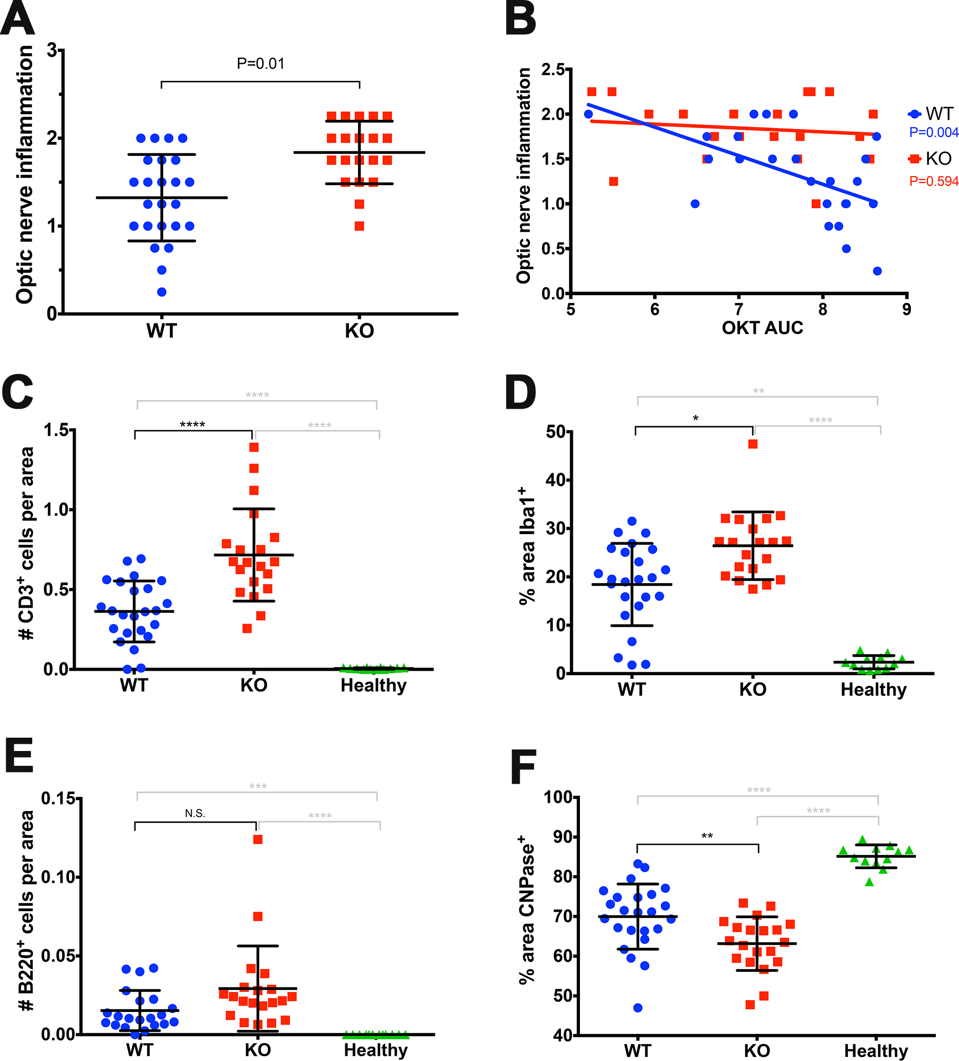
 Figure 4 of
Larabee, Mol Vis 2016; 22:1503-1513.
Figure 4 of
Larabee, Mol Vis 2016; 22:1503-1513.