Figure 2. Nrf2-deficient and wild-type mice exhibit different immune responses to EAE despite comparable immune cell profiles before
EAE challenge. Flow cytometric analysis of cells recovered from the thymus (A), bone marrow (B), and spleen (C) of healthy, non-experimental autoimmune encephalomyelitis (EAE) wild-type (WT; blue, left) and nuclear factor-E2-related
factor (Nrf2) knockout (KO; red, right) mice. The antibodies used detected CD4+ T-cells, CD8+ T-cells, CD4+CD8+ T-cells (A), Gr-1+ granulocytes (B, C), Iba1+ macrophages (B, C), and CD19+ B-cells (B, C). Whiskers indicate min/max. n = 6 (three male mice, three female mice) per group. Mann–Whitney analysis. D–F: ELISA analysis of WT (blue circles) and Nrf2 KO (red squares) EAE spleens to determine the interleukin-6 (IL-6; D), IL-17 (E), and interferon-gamma (IFN-γ; F) cytokine levels. n = 10–12. Mann-Whitney was used for statistical analysis.
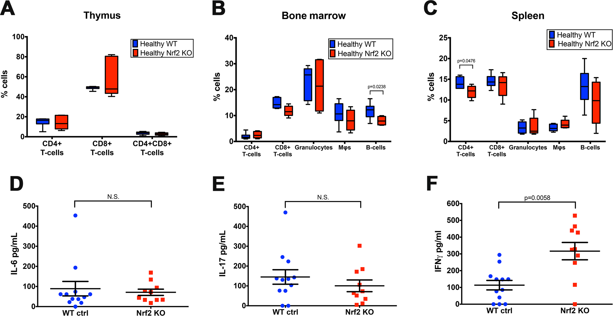
 Figure 2 of
Larabee, Mol Vis 2016; 22:1503-1513.
Figure 2 of
Larabee, Mol Vis 2016; 22:1503-1513.