Figure 3. Wide-field fundus autofluorescence images of the patients with cone or cone-rod dystrophy carrying pathogenic mutations. Fundus
autofluorescence was imaged with Optos200Tx (Optos, Dunfermline, UK) except K6140 (HRA2; Heidelberg Engineering, Heidelberg,
Germany). Most of the examinations for K6205, the mother of K6073, were performed at another institution and were not available.
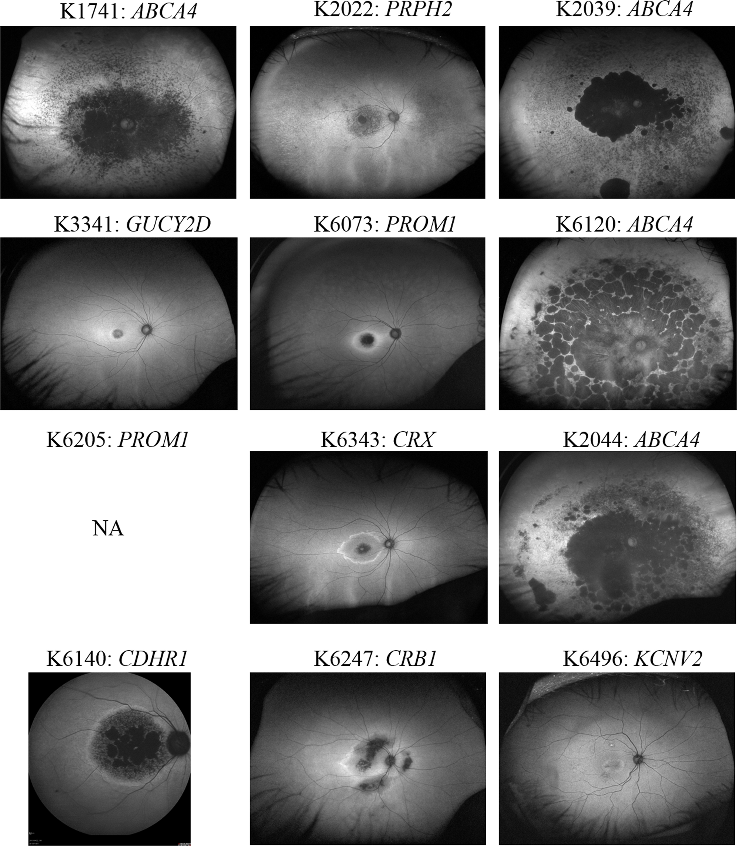
 Figure 3 of
Oishi, Mol Vis 2016; 22:150-xxx.
Figure 3 of
Oishi, Mol Vis 2016; 22:150-xxx.