Figure 5. Effect of CDECM on oxidative stress in hConECs and hPECs. A: Intracellular reactive oxygen species (ROS) were detected using 2′,7′-dichlorofluorescein diacetate (DCFH-DA), a ROS-sensitive
fluorescent dye. The stained cells were counterstained with 4′,6-diamidino-2-phenylindole (DAPI) and viewed under an automatic
fluorescence in situ hybridization (FISH) imager. Scale bar = 50 μm. The cell lysates were immunostained for Nox2 and p47phox
(B). The graphs show the densitometry quantification of Nox2 (C) and p47phox (D). β-actin was used as the control. Values are mean ± standard deviation (SD; n = 5). Statistical significance is indicated
as **p<0.01. E: The cell lysates were immunostained for heme oxygenase-1 (HO-1) and nuclear factor erythroid-2 related factor 2 (Nrf2).
The graphs show the densitometry quantification of HO-1 (F) and Nrf2 (G). β-actin and Lamin B were used as the control for the whole cell fraction and the nucleus fraction, respectively. Values
are mean ± SD (n = 5). Statistical significance is indicated as *p<0.05 and **p<0.01.
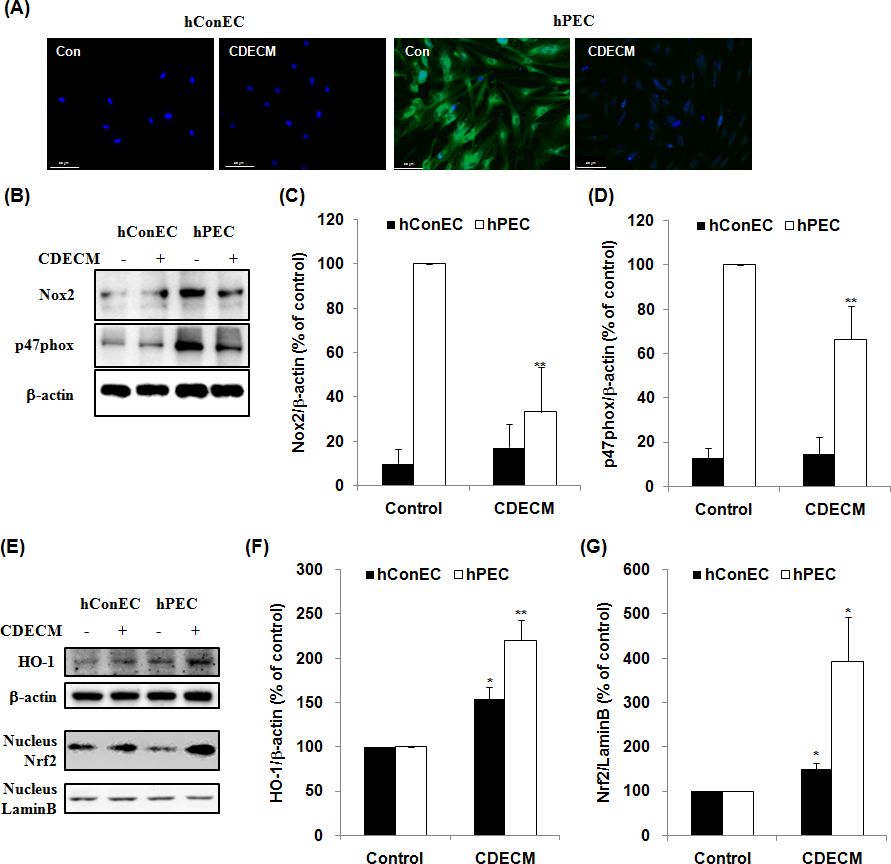
 Figure 5 of
Lee, Mol Vis 2016; 22:1490-1502.
Figure 5 of
Lee, Mol Vis 2016; 22:1490-1502.