Figure 4. Effect of CDECM on angiogenesis and inflammation in hConECs and hPECs. A: The cells were immunostained for tumor necrosis factor-α (TNF-α), antivascular cellular adhesion molecule 1 (VCAM-1), cyclooxygenase-2
(Cox2), vascular endothelial growth factor (VEGF), and cluster of differentiation 31 (CD31). Images of the membranes were
photographed with the Fusion FX image acquisition system. The graphs show the densitometry quantification of TNF-α (B), VCAM-1 (C), Cox2 (D), VEGF (E), and CD31 (F), compared with the untreated human pterygium epithelial cells (hPECs). β-actin was used as the control. Values are mean
± standard deviation (SD; n = 5). Statistical significance is indicated as *p<0.05, **p<0.01, and ***p<0.001 (B). The levels of interleukin 1 beta (IL-1β) (G), IL-6 (H), and prostaglandin E2 (PGE2) (I) production were measured with enzyme-linked immunosorbent assay (ELISA). Data are expressed as the means ± SD (n = 3). Statistical
significance is indicated as ***p<0.001.
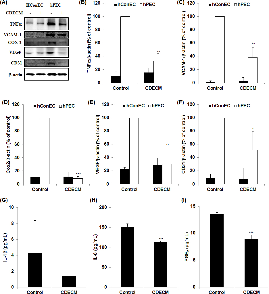
 Figure 4 of
Lee, Mol Vis 2016; 22:1490-1502.
Figure 4 of
Lee, Mol Vis 2016; 22:1490-1502.