Figure 8. Chx10 colocalized with CCND1 before BC differentiation. The developing (A–C) PN1 and (D–F) PN3 control retinas double labeled with antibodies against Chx10 (green: A and D) and CCND1 (red: B and E), and colabeling was examined in the merged images (yellow: C and F). A: In the PN1 controls, Chx10-IR was diffusely located throughout the retina in the cytosolic compartment. B: CCND1, a cell cycle protein, was localized in the nucleus of the proliferating cells in the VZ. C:Chx10-IR colocalized with CCND1-IR in the VZ. D: In the PN5 control retinas, Chx10-IR was localized in the somas in the INL and the cytosol of cells in the ganglion cell layer. The shape of the Chx10-IR in the INL cells changed from spindly to compact and round. E: CCND1 localized to spindly proliferating cells in the INL. F:Chx10 did not colabel with CCND1 in the INL. Scale bar = 40 μm. CCND1 = cyclin D1; BC = bipolar cell; PN = postnatal; VZ = ventricular
zone; INL = inner nuclear layer.
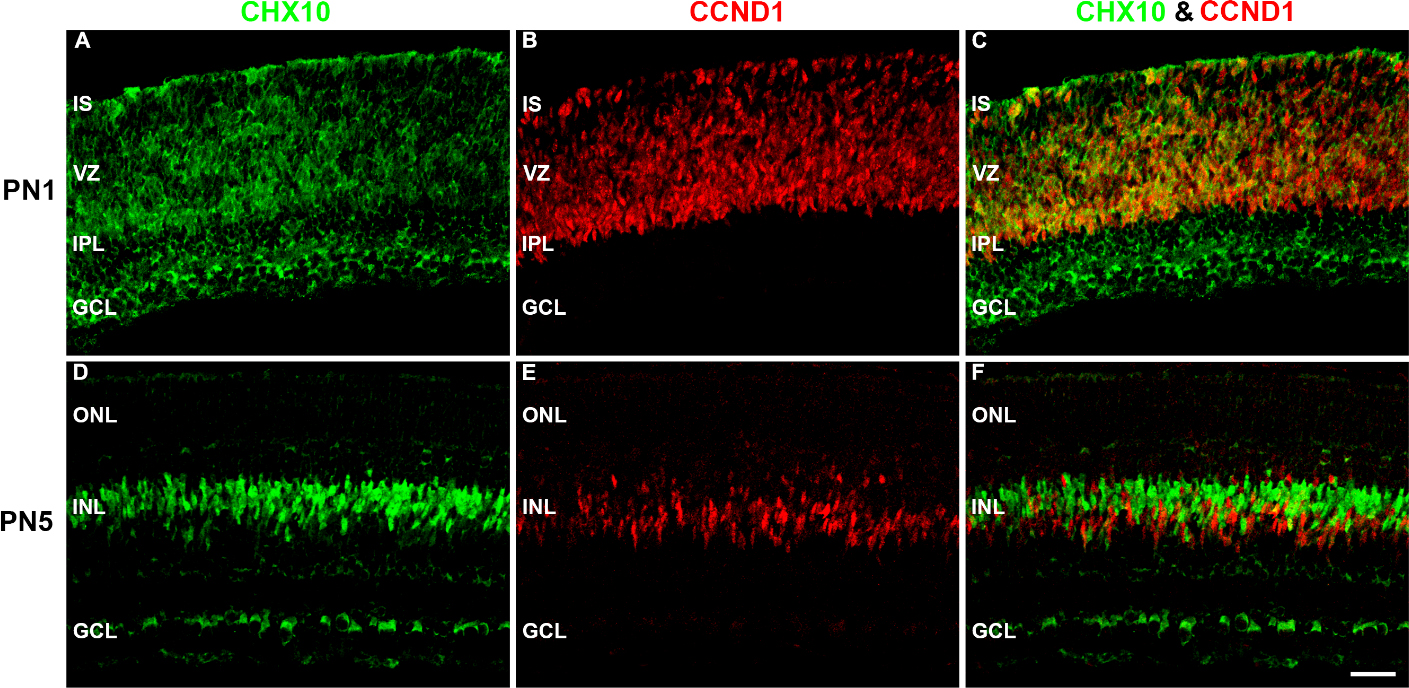
 Figure 8 of
Chaney, Mol Vis 2016; 22:1468-1489.
Figure 8 of
Chaney, Mol Vis 2016; 22:1468-1489.