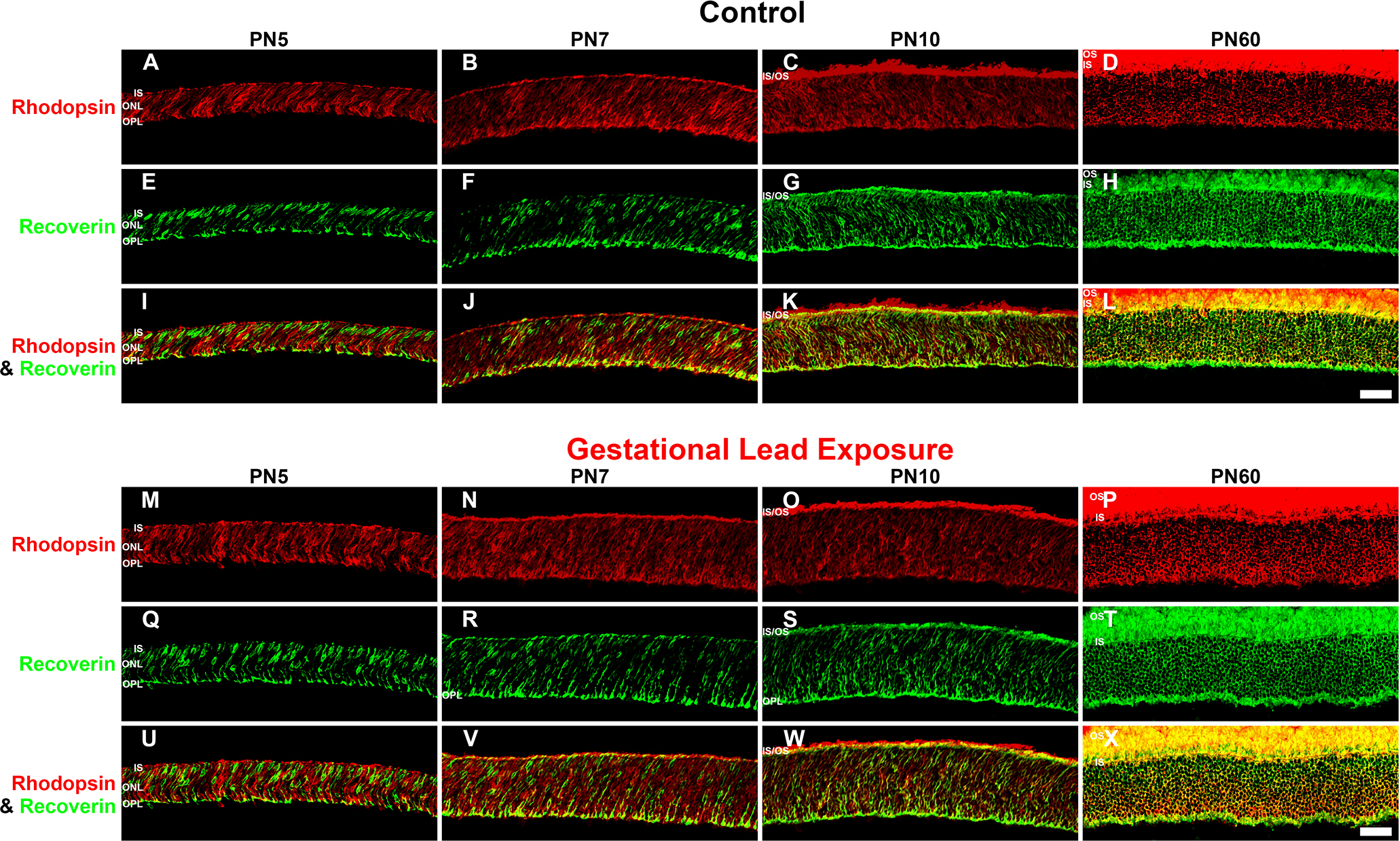
Figure 3. GLE delayed rhodopsin-IR, but not recoverin-IR, in developing retinas (PN5-PN60). The developing retinas at PN5-PN60 from
(
A–L) the control and (
M–X) GLE mice were double labeled with antibodies against rhodopsin (red:
A–D and
M–P) and recoverin (green:
E–H and
Q–T), and colabeling was examined in merged images (yellow:
I–L and
U–X). In the control retinas at (
A,
E, and
I) PN5 and (
B,
F, and
J) PN7, the ISs, ONL, and OPL were colabeled with rhodopsin and recoverin, which increased at PN7.
C,
G, and
K: At PN10, the OS expressed rhodopsin-IR, and extensive colabeling with recoverin occurred in the ISs, ONL, and OPL.
D,
H, and
L: In young adult control retinas (PN60), the OSs were intensely rhodopsin-IR, and there was extensive labeling in the ISs,
ONL, and OPL. In the distal OPL, the smaller rod spherules [
24] were colabeled (yellow pixels), whereas the larger cone pedicles in the proximal OPL [
24] were only recoverin-IR (green pixels). In the GLE retinas at (
M,
Q,
and U) PN5 and (
N,
R, and
V) PN7, the ISs, ONL, and OPL were rhodopsin-IR and recoverin-IR, and an increased amount of colabeling was seen in all the
layers. Relative to the age-matched controls, the ONL thickness increased.
O,
S, and
W: At PN10, OSs were rhodopsin-IR, and extensive colabeling with recoverin occurred in the ISs, ONL, and OPL. Relative to the
age-matched controls, the ONL thickness increased.
P,
T, and
X: In the PN60 GLE retinas, the OSs, ONL, and OPL were intensely rhodopsin-IR and almost completely colabeled with recoverin.
Relative to the age-matched controls, the ONL and OPL thickness increased, and the number of rod spherules increased as described
[
21]. Scale bar = 40 μm. GLE = Gestational lead exposure; IR = immunoreactivity; PN = postnatal; IS = inner segment; OS = outer
segment; ONL = outer nuclear layer; OPL = outer plexiform layer.
 Figure 3 of
Chaney, Mol Vis 2016; 22:1468-1489.
Figure 3 of
Chaney, Mol Vis 2016; 22:1468-1489.