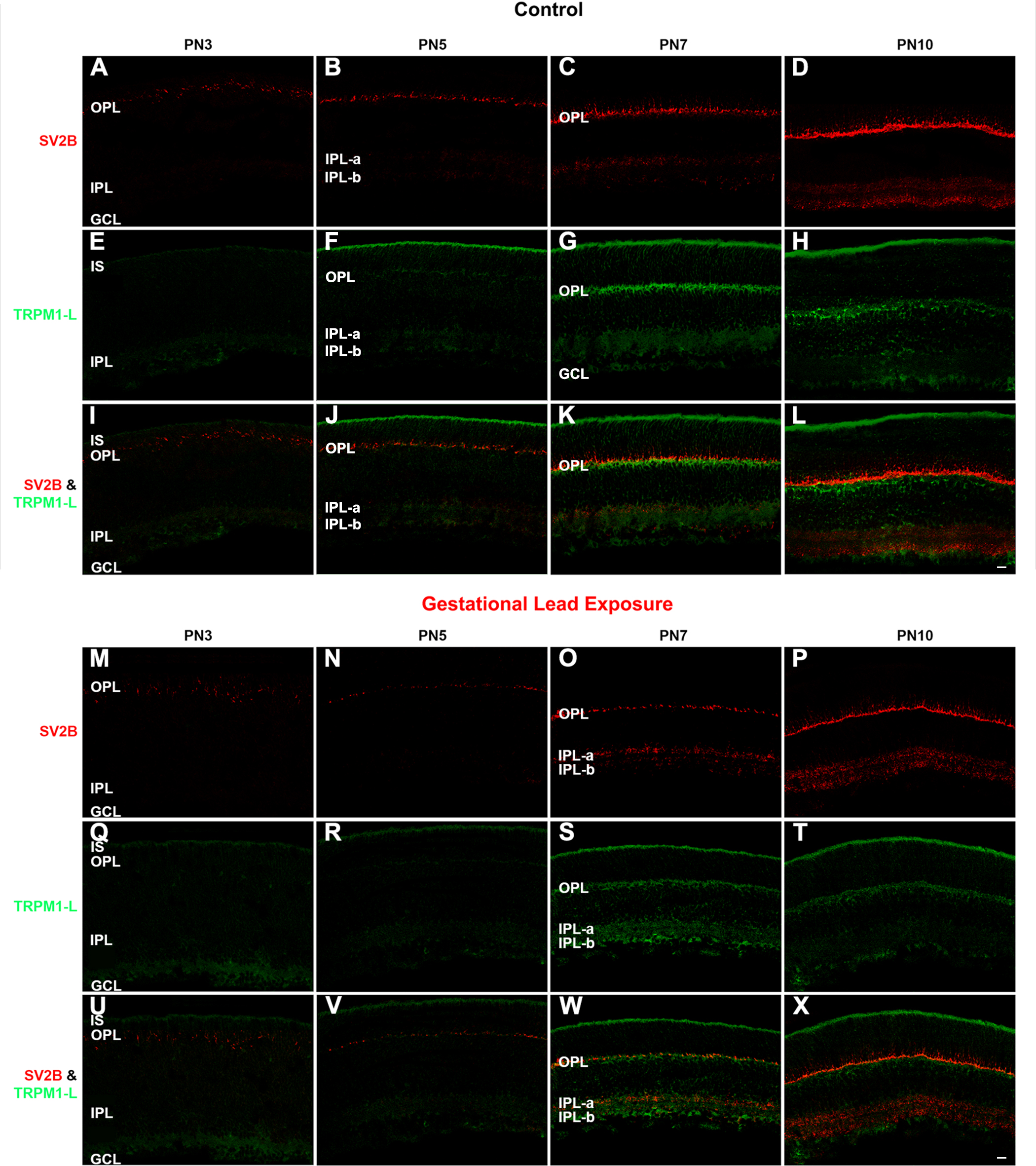
Figure 11. GLE delayed SV2B- and TRPM1-IR in the developing plexiform layers. The developing retinas from (
A–L) the control and (
M–X) GLE mice were double labeled with antibodies against SV2B (red:
A–D and
M–P) and TRPM1-L (green:
E–H and
Q–T), and colabeling was examined in the merged images (yellow:
I–L and
U–X).
A–B: In the PN3 and PN5 controls, SV2B was visible in the OPL. In the PN5 retinas, punctate SV2B-IR was visible in the IPL.
C: At PN7, SV2B-IR occurred more intensely in the OPL and IPL, with higher intensity in IPL-a than IPL-b.
D: By PN10, SV2B-IR laminated in the OPL such that there were discrete puncta in the distal (rod spherules) and clumps in the
proximal (cone pedicles) layers. The IPL laminated into IPL-a, and IPL-b separated by a layer that was not labeled.
E: In the PN3 controls, TRPM1-L diffusely labeled the ISs, IPL, and GCL.
F: By PN5, TRPM1-L intensely labeled the IS and weakly labeled the developing OPL.
G: At PN7 TRPM1-L-IR intensified in the ISs, OPL, and IPL. Furthermore, the TRPM1-L labeled the distal somas of the BCs right
below the OPL.
H: This pattern of TRPM1-L-IR persisted at PN10.
I–J: No colabeling was observed at PN3 and PN5.
K–L: In the OPLs of the PN7 and PN10 controls, SV2B labeled just above TRPM1-L-IR with little to no overlap. No colabel was observed
in the IPL.
M: In the PN3 GLE retinas, SV2B-IR puncta in the developing OPL was not uniformly localized as in the age-matched controls.
N–O: The OPLs of the PN5 and PN7 GLE retinas were less mature than those in the age-matched controls. Little SV2B-IR was observed
in the IPL of the PN5 GLE retinas. However, by PN7, SV2B labeled the IPL; IPL-a labeled more strongly than IPL-b.
P: In the PN10 GLE retinas, SV2B-IR in the OPL and IPL increased; however, IPL-a and IPL-b did not show differential labeling
as in the control.
Q–T: TRPM1-L-IR in the GLE retinas was similar to the controls, except at PN7, where the OPL was not labeled as brightly.
U–V: SV2B and TRPM1-L did not colocalize at PN3 and PN5 in the GLE retinas as in the controls.
W–X: The PN7 and PN10 GLE retinas differed from the controls; at PN7, SV2B labeled in the same plane as, rather than above, TRPM1-L.
At PN10, SV2B colocalized with TRPM1-L more than in the controls. Thus, there was a two-day delay in the appearance of SV2B-IR
and TRPM1 in the GLE retinas (
Table 3). Scale bar = 40 μm. GLE = Gestational lead exposure; SV2B = synaptic vesicle protein 2B; TRPM1-L = transient receptor potential
M1-long; IR = immunoreactivity; OPL outer plexiform layer; IPL = inner plexiform layer; PN = postnatal; IS = inner segment;
GCL = ganglion cell layer.
 Figure 11 of
Chaney, Mol Vis 2016; 22:1468-1489.
Figure 11 of
Chaney, Mol Vis 2016; 22:1468-1489.