Figure 4. Developmental expression of cone transducin γ protein in the embryonic and postnatal retina. Immunohistochemical staining
was performed on coronal sections starting at (A) E10.5 (before cone birth) through (I) P21. Beginning at E13.5, cone transducin γ expression is observed in cells along the ventricular surface (arrows in D), and this expression continues through all ages tested. Scale bars = 100 µm.
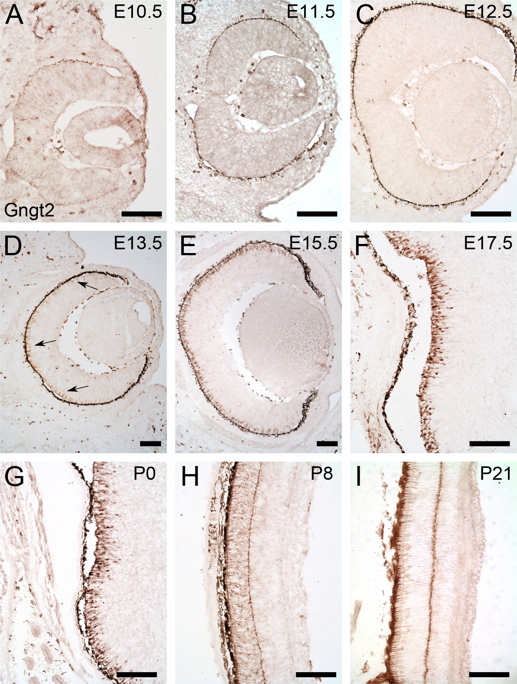
 Figure 4 of
Rodgers, Mol Vis 2016; 22:1455-1467.
Figure 4 of
Rodgers, Mol Vis 2016; 22:1455-1467.