Figure 3. Colocalization of phosducin with known photoreceptor markers in the embryonic retina. A: Fluorescent imaging of coronal sections of mouse retinas at E15.5 shows the colocalization of phosducin and Otx2, a photoreceptor
marker, along the ventricular surface. Otx2 shows RPE labeling (arrowheads) in addition to photoreceptor labeling. Otx2-positive,
Pdc-negative migrating cells are also seen interior to the ventricular surface (arrows). B: Immunofluorescent imaging of the retinal sections at E17.5 for phosducin and the cone marker, Rxrγ, shows consistent colocalization
along the ventricular surface of the retina. Scale bars = 50 µm.
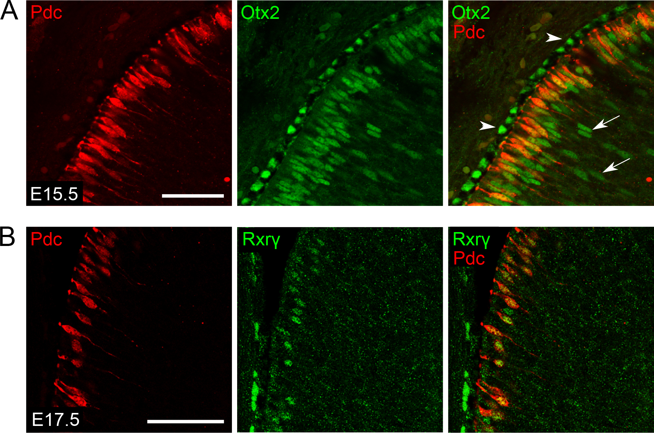
 Figure 3 of
Rodgers, Mol Vis 2016; 22:1455-1467.
Figure 3 of
Rodgers, Mol Vis 2016; 22:1455-1467.