Figure 2. Comparison of PdcKO and control retinas. Immunofluorescence was performed on coronal sections of control and phosducin knockout
(PdcKO) mice at ages E13.5 (A) using anti-Pdc and anti-Otx2 antibodies and P0 (B) using anti-Pdc and anti-cone transducin γ (Gngt2) antibodies. Pdc expression is seen in the control retinas and is absent
in the PdcKO retinas, whereas Otx2 and Gngt2 are expressed in the control and PdcKO retinas. Scale bars = 100 µm.
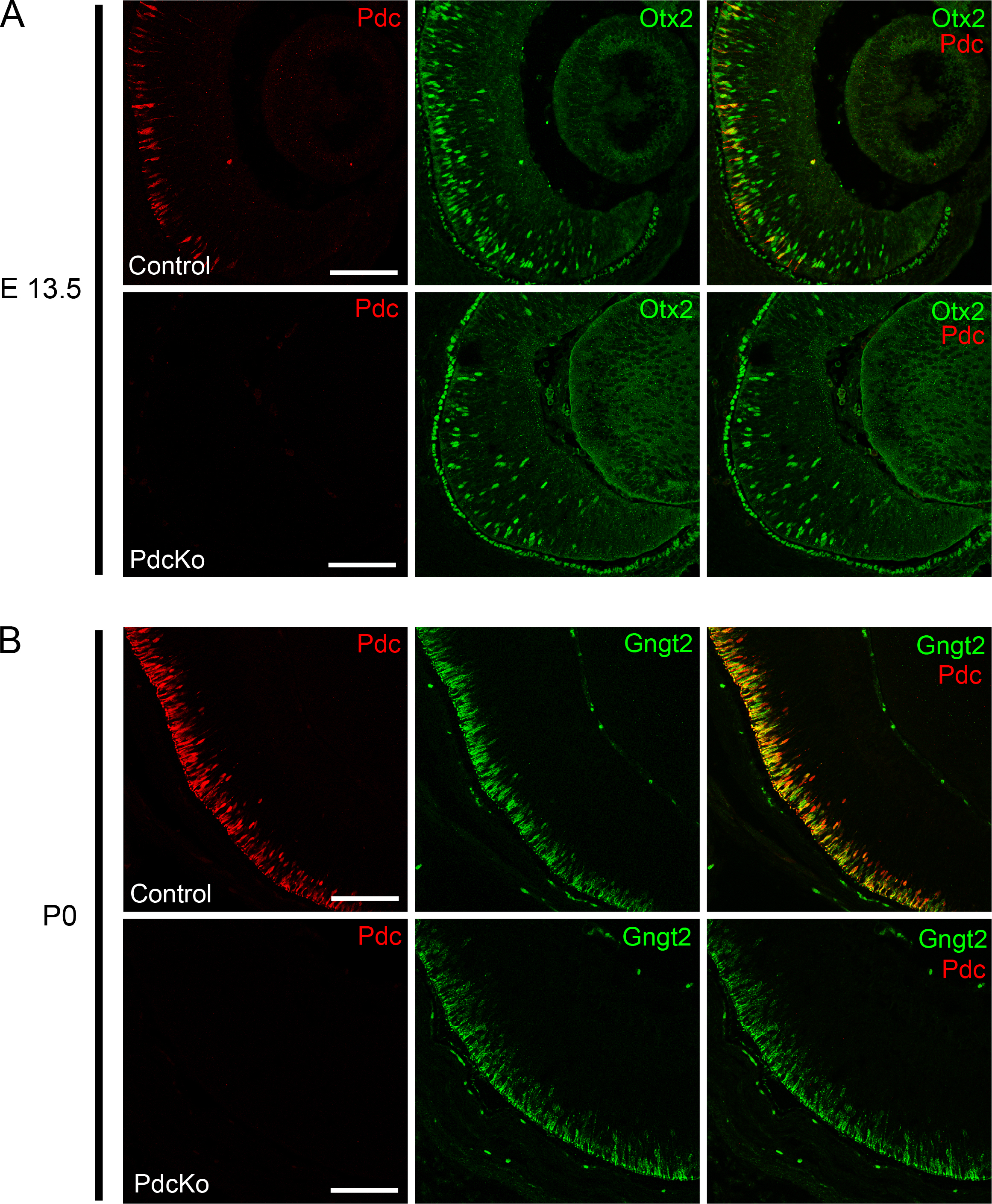
 Figure 2 of
Rodgers, Mol Vis 2016; 22:1455-1467.
Figure 2 of
Rodgers, Mol Vis 2016; 22:1455-1467.