Figure 3. Immunofluorescence analysis of the human retrolaminar optic nerve. MMP19 immunolabeling is present in the peripheral (A, D, green) and central regions (G, J, green) of the optic nerve, as well as collagen IV (B, H, red) and UEA-I lectin labeling (E, K, red). The merged images show colocalization between MMP19 and collagen IV (C, I), while MMP19 and UEA-I do not overlap except in large vessel walls (F, L). Sections were counterstained with 4’6-diamidino-2-phenylindole (DAPI; C–L). Scale bar = 100 µm. d, dura mater, a, arachnoid mater; p, pia mater.
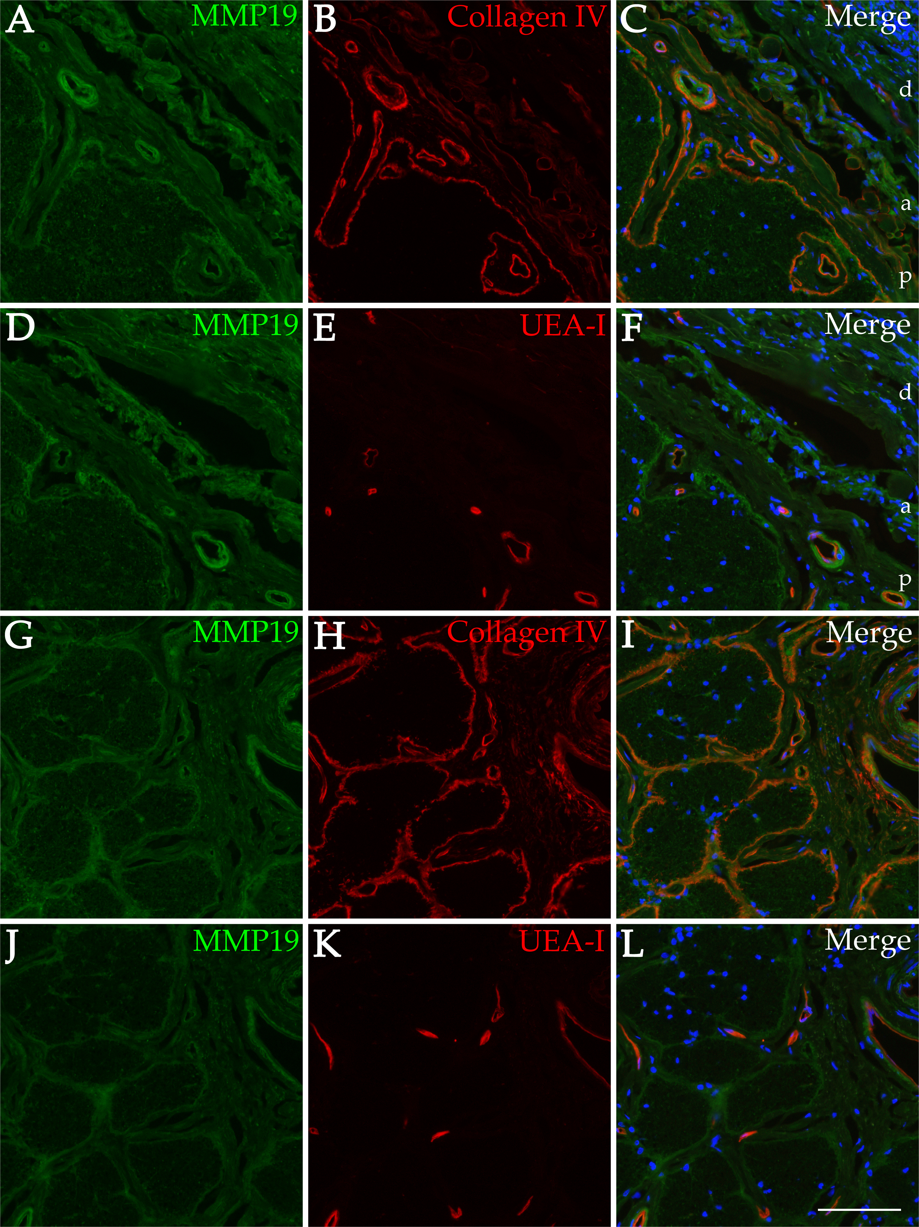
 Figure 3 of
Chirco, Mol Vis 2016; 22:1429-1436.
Figure 3 of
Chirco, Mol Vis 2016; 22:1429-1436.