Figure 2. Immunohistochemistry of the human retrolaminar optic nerve. Anti-MMP19 antibody was used to label a central region (A) and a peripheral region (B) of the retrolaminar optic nerve (purple labeling indicates the reaction product). C, D: The anti-MMP19 antibody was preincubated with full-length MMP19 protein before immunolabeling. E: A grayscale, negative image of the MMP19 immunolabeling for the entire width of the optic nerve is presented. Scale bar
(D) = 100 µm; scale bar (E) =500 µm. a, arachnoid mater; p, pia mater.
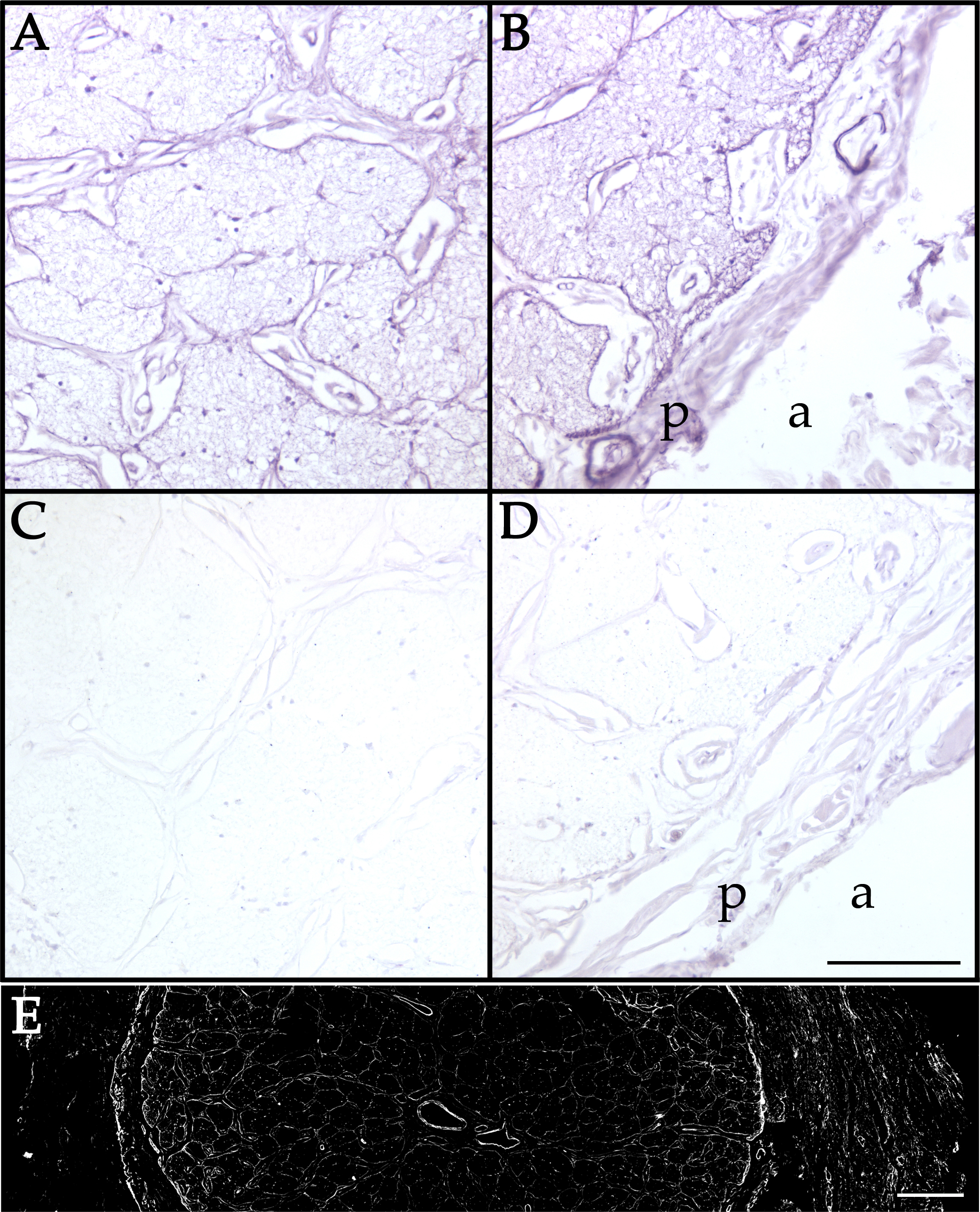
 Figure 2 of
Chirco, Mol Vis 2016; 22:1429-1436.
Figure 2 of
Chirco, Mol Vis 2016; 22:1429-1436.