Figure 3. vsx1 and vsx2 are expressed in the INL of the maturing Xenopus laevis retina. A–H: Expression of vsx1 (A–D) and vsx2 (E–H) at st 38, 41, 45, and 50 (as indicated). Brackets indicate the ciliary marginal zone (CMZ). I–L: Overlapping expression of vsx1 and vsx2 decreases as the retina matures. Double in situ hybridization was performed on sections prepared from paraffin-embedded tadpoles
using differently labeled probes for vsx1 (red) and vsx2 (blue). I, inner nuclear layer; G, ganglion cell layer; L, lens. Scale bars = 1 µM. Scale bar in A applies to B, C, E, F, and G; scale bar in D applies to H.
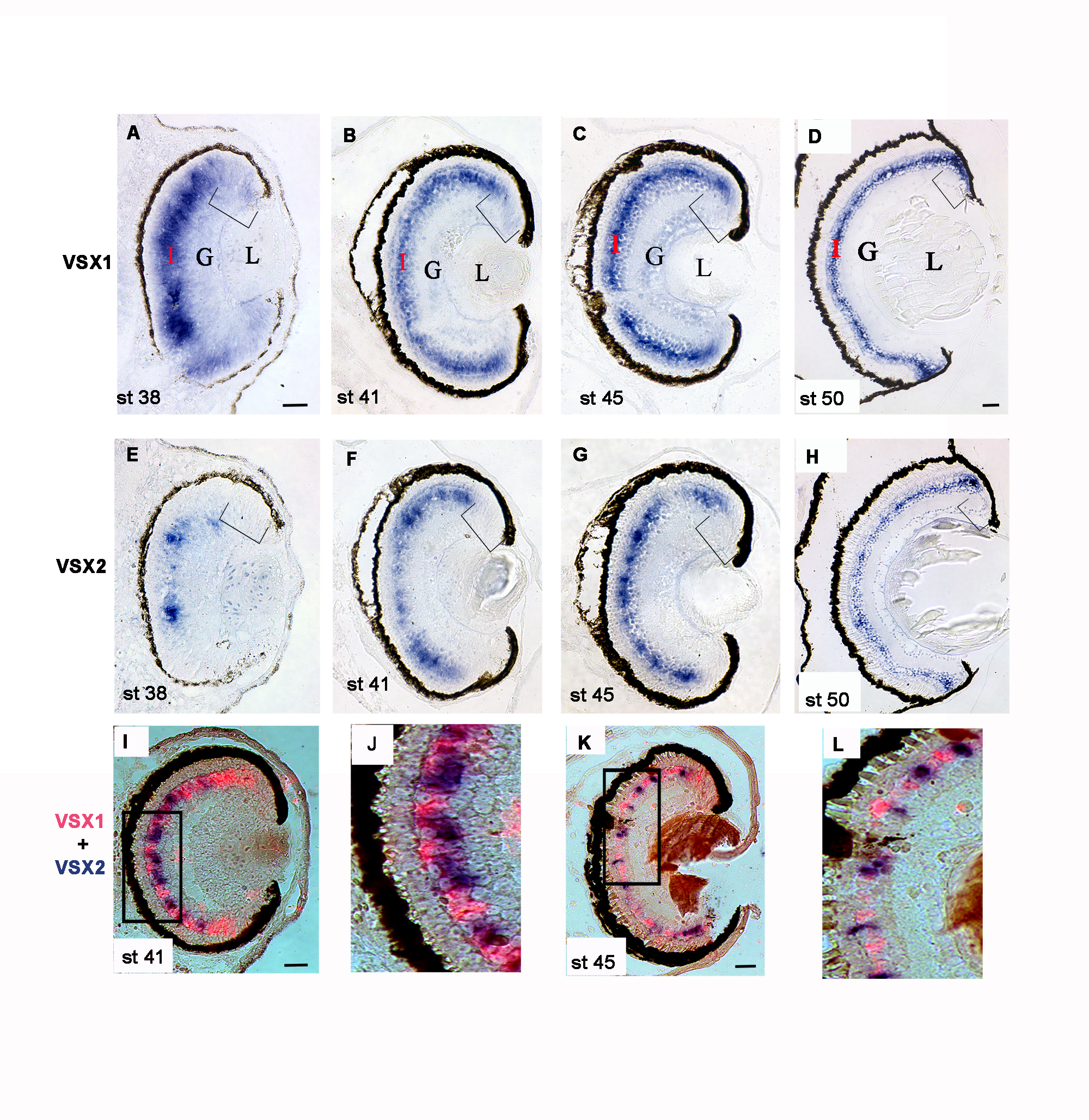
 Figure 3 of
Pan, Mol Vis 2016; 22:1421-1428.
Figure 3 of
Pan, Mol Vis 2016; 22:1421-1428.