Figure 1. Identification of Xenopus laevis Vsx1 and Vsx2. A: Predicted protein sequences of X. laevis Vsx1 and Vsx2. Sequences were aligned using ClustalW. The black and gray backgrounds indicate amino acid identity and similarity,
respectively. The highly conserved octapeptide motif (OP; box), homeodomain (dashed line), and CVC domain (double underline)
are indicated. The orthopedia-aristaless-Rx (OAR; Vsx2) and Rinx-Vsx1 (RV; Vsx1) domains are also indicated. The triple underline
indicates the region of Vsx2 lacking from the short form. B: Phylogenetic representation of ClustalW alignment of the Vsx1 and Vsx2 amino acid sequences from X. laevis, zebrafish, chicken, and mouse, Caenorhabditis elegans Ceh-10, and X. laevis Rx-L.
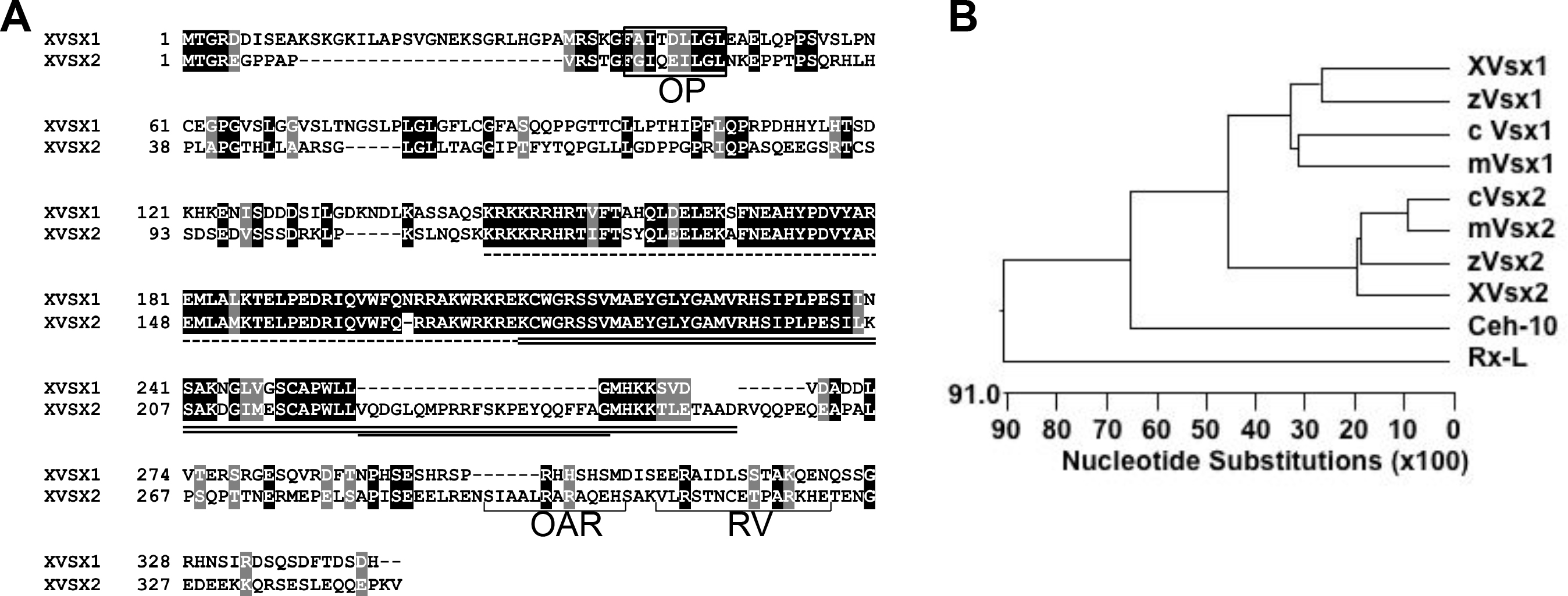
 Figure 1 of
Pan, Mol Vis 2016; 22:1421-1428.
Figure 1 of
Pan, Mol Vis 2016; 22:1421-1428.