Figure 6. Displaced cells and electroporation of a stage 12 optic vesicle. A–B: Fluorescence micrograph of a sectioned retina from an embryonic day 14 (E14) embryo electroporated with the RXRγ208 Cre-LoxP
piggyBac constructs in ovo at stage 28, showing a displaced photoreceptor (PR; arrow) and a displaced horizontal cell (HC;
arrow). C: Fluorescence micrograph of a sectioned retina from an E14 embryo electroporated with the RXRγ208 Cre-LoxP piggyBac constructs
in ovo at stage 12.
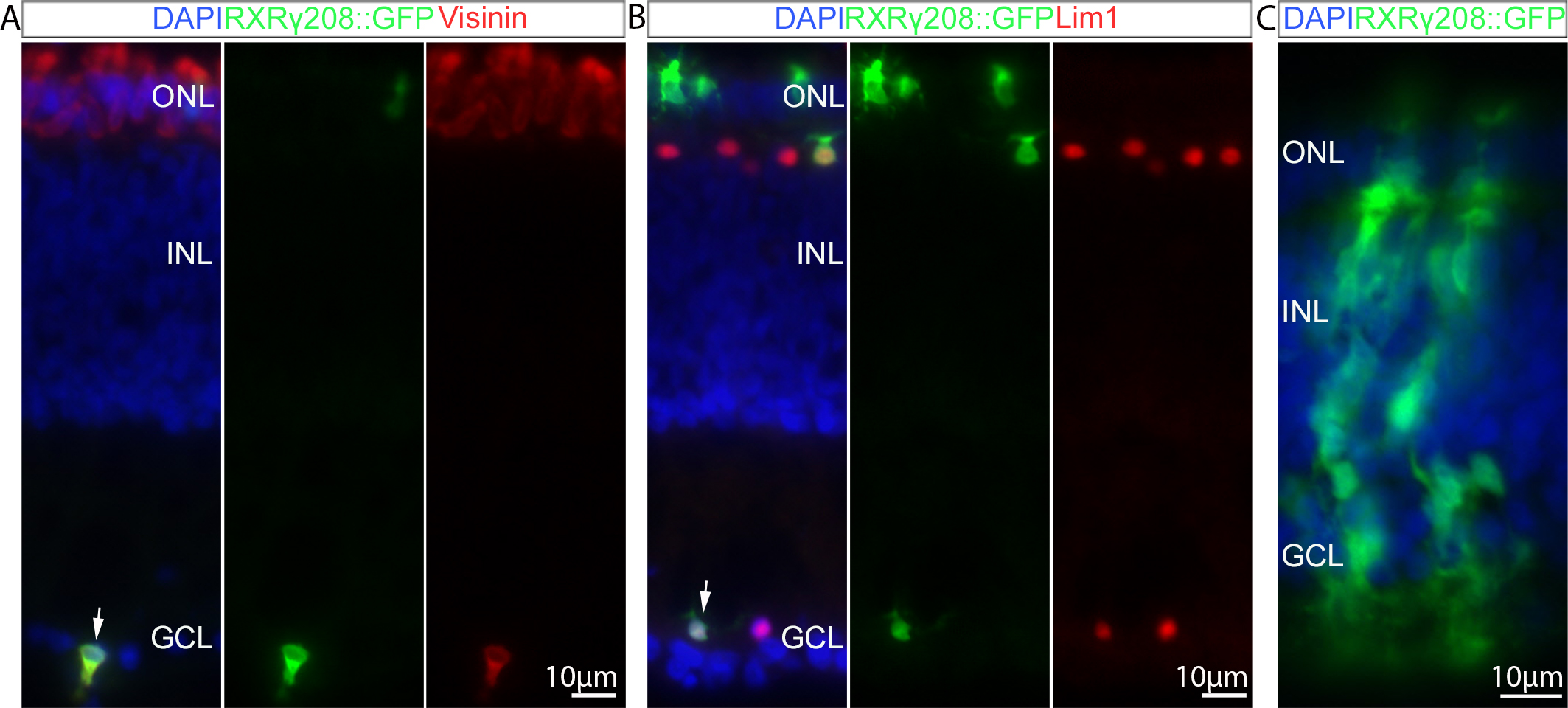
 Figure 6 of
Blixt, Mol Vis 2016; 22:1405-1420.
Figure 6 of
Blixt, Mol Vis 2016; 22:1405-1420.