Figure 4. Activity of the RXRγ208 sequence produced specific reporter gene expression. A: Z-stacked confocal fluorescence micrograph of a flatmounted retina from a stage 40 embryo electroporated with the RXRγ208
Cre-LoxP piggyBac constructs in ovo at stage 25. B: Fluorescence micrograph of a retina from a stage 40 embryo electroporated with the RXRγ208 Cre-LoxP piggyBac constructs
in ovo at stage 28. C: Bar graph showing the distribution of green fluorescent protein–positive (GFP+) cells in sectioned retinas from embryos
electroporated with the RXRγ208 Cre-LoxP piggyBac constructs in ovo at stages 22, 25, and 28 (mean ± standard deviation, SDSD,
n = 4 per stage). D: Fluorescence micrograph of a retina showing a pair of photoreceptors (PRs; arrow heads) and a pair with a horizontal cell
(HC) and a PR (arrows). St = stage, ONL = outer nuclear layer, INL = inner nuclear layer, GCL = ganglion cell layer.
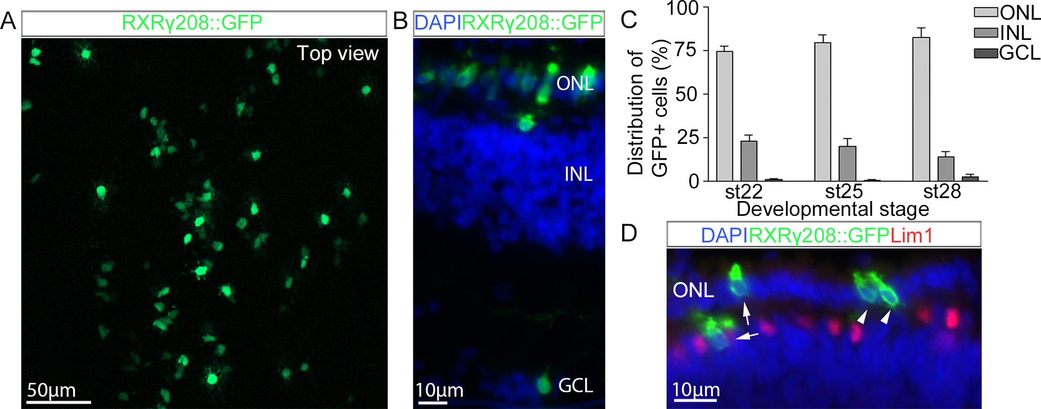
 Figure 4 of
Blixt, Mol Vis 2016; 22:1405-1420.
Figure 4 of
Blixt, Mol Vis 2016; 22:1405-1420.