Figure 2. Evaluation of the Cre-LoxP piggyBac transposon system. A: Fluorescence micrographs of chicken fibroblast DF1-cells electroporated with the piggyBac constructs and a red fluorescence
protein (RFP) control, and analyzed 3, 7, or 14 days post-treatment. B´: Diagram of the RFP-expressing control construct. B´´: Diagrams of the green fluorescent protein (GFP) reporter gene constructs used for integrations into the host cell genome.
C: Micrographs of the whole retina and of the section from a stage 34 retina electroporated with the Stacia3-cytomegalovirus
(CMV), CAG-PBase, and pB-RAGE-green fluorescent protein (GFP) constructs in ovo at stage 12. Asterisk (*) indicates the optic
nerve exit. D: Fluorescence micrographs of stage 36 retinas that were electroporated with the Cre-LoxP piggyBac constructs in ovo at stages
22, 25, and 28. St = stage, ONL = outer nuclear layer, INL = inner nuclear layer, GCL = ganglion cell layer.
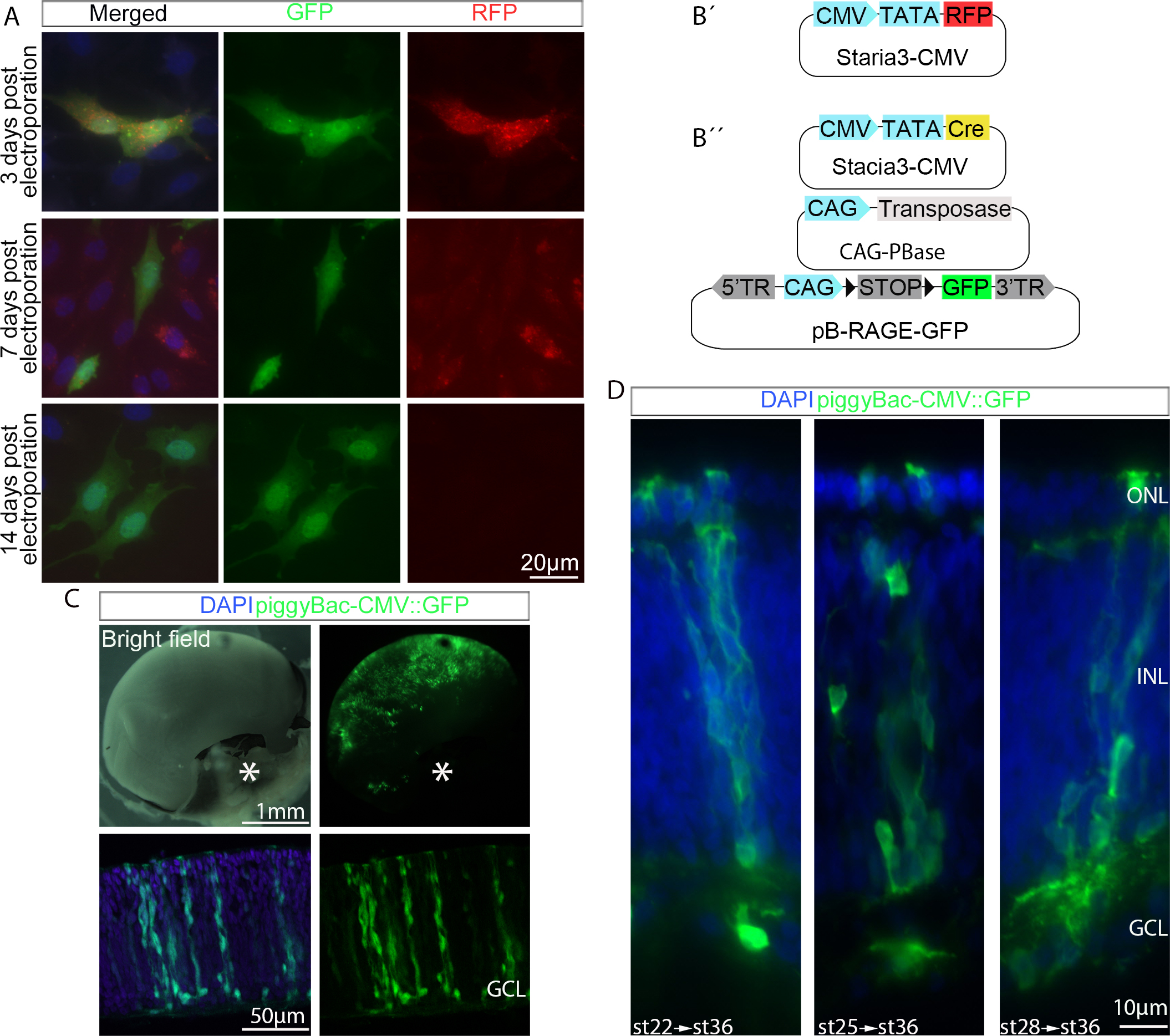
 Figure 2 of
Blixt, Mol Vis 2016; 22:1405-1420.
Figure 2 of
Blixt, Mol Vis 2016; 22:1405-1420.