Figure 5. LDLR localization and LDL uptake in serum-deprived and -starved ARPE-19 cells. A: Immunofluorescence of cells in serum-free medium (SFM) from day 0–7. Low-density lipoprotein receptor (LDLR; green) is primarily
intracellular at day 0, but by day 7 in SFM, more signal appears localized to the cell surface. Images are 40X with scale
bar indicating 20 μM, except the final panel with 100X magnification for day 7. Scale bar=5 μM. Nuclei are stained with 4',6-diamidino-2-phenylindole
(DAPI; blue). B: Uptake of labeled LDL by ARPE-19 cells in SFM. ARPE-19 cells in SFM were incubated with LDL-DyLight 549, washed and fixed,
and the nuclei were stained with DAPI (blue). Confocal images show intracellular LDL-DyLight 549 in red. Scale bars=20 µm.
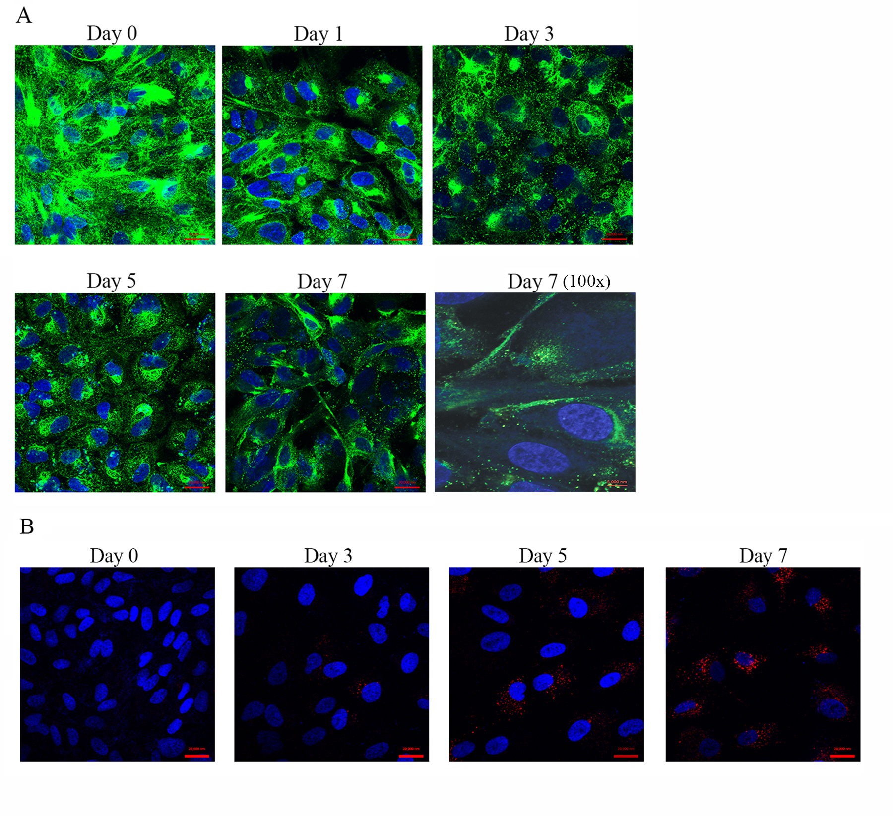
 Figure 5 of
Mishra, Mol Vis 2016; 22:1387-1404.
Figure 5 of
Mishra, Mol Vis 2016; 22:1387-1404.