Figure 9. Phase contrast micrographs of outgrowths from limbal–peripheral biopsies obtained from donors and the contralateral regenerated
corneas. A and B: Outgrowths from a cornea regenerated with a primary-generation explant culture (top) and from its contralateral donor eye
(bottom). C–F: Equivalent micrograph pair from a secondary outgrowth-based regeneration (C) and from three tertiary outgrowth-based regenerations (D–F). The regularly distributed white puncta on these images arises from reflections of the 0.4 μm pore of the permeable substratum.
The scale bars equal 250 μm in A and 50 μm in B through F.
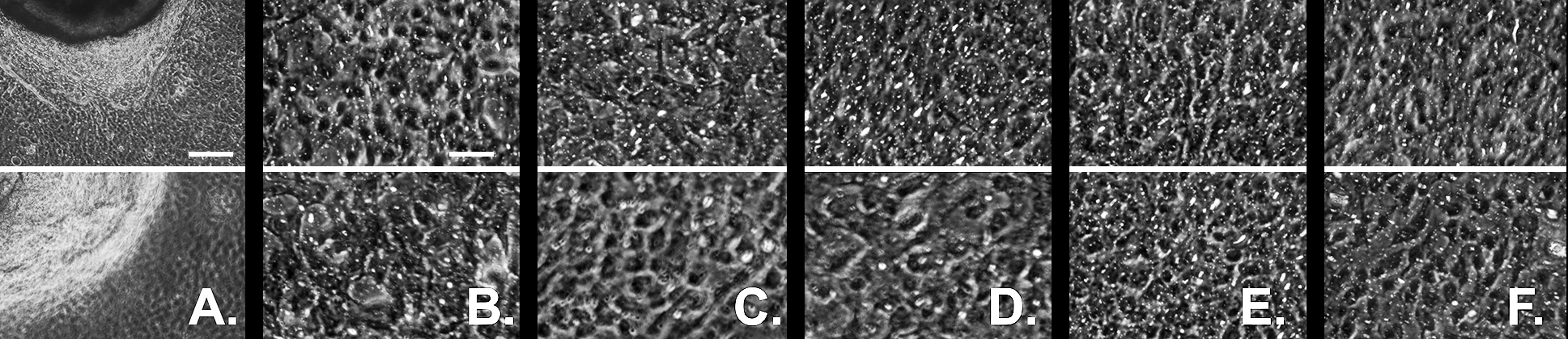
 Figure 9 of
Selver, Mol Vis 2016; 22:138-149.
Figure 9 of
Selver, Mol Vis 2016; 22:138-149.