Figure 6. Histological and p63 immunochemical features of four treated corneas grafted with tertiary-generation limbal explant outgrowths.
Hematoxylin and eosin (H&E) composites of the full cornea (A) or large areas of it (B and C) are shown. Unframed inserts in B and C show magnified H&E details of the limbal–peripheral and corneal zone. Framed inserts show p63 staining of respective adjacent
sections at arbitrary magnifications.
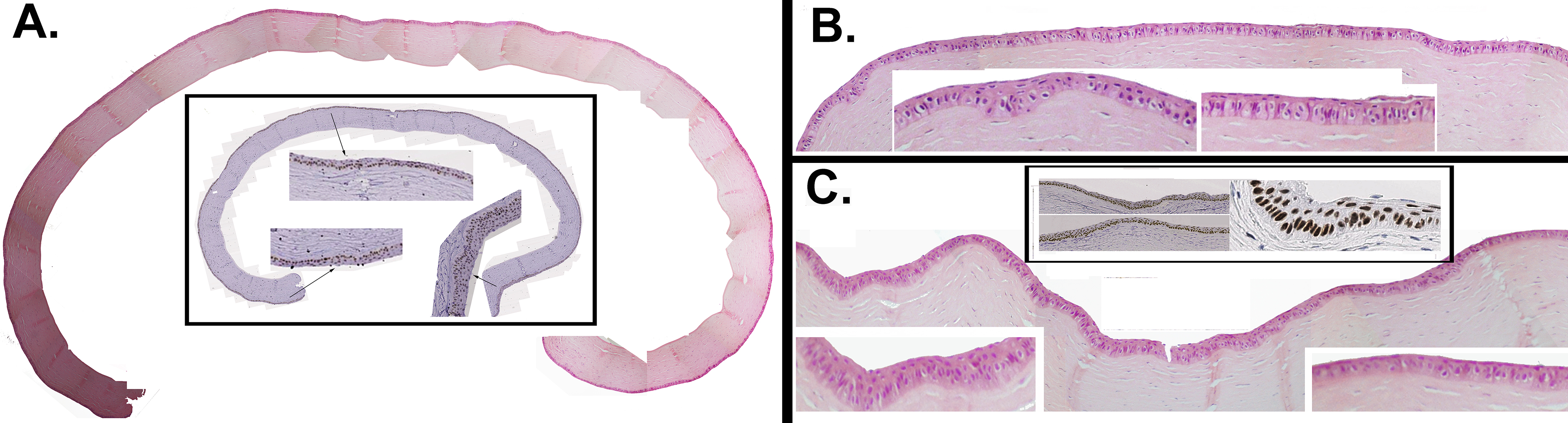
 Figure 6 of
Selver, Mol Vis 2016; 22:138-149.
Figure 6 of
Selver, Mol Vis 2016; 22:138-149.