Figure 5. Histological and immunochemical details of the limbal and corneal zone of the control ocular surface and ocular surfaces grafted
with primary outgrowths following radical limbal excision and corneal epithelial removal. A–E, L, and M: Donor cornea. The area selected was not subjected to surgical biopsy excision. A–C: Hematoxylin and eosin (H&E) stain at three magnifications. Note the loose stroma defining the limbal papilla. D and E: p63 stain of the limbal and central corneal zone. Note that p63 is strongly expressed in the nuclei of most cells in both
zones, including some suprabasal nuclei. L and M: Krt3 stain in the limbus and the central cornea. Note the pronounced limbal rete ridge appearance in the limbus and the
absence of Krt3 in the basal cell layer there. F–K, N, and O: Ocular surface grafted with a primary-generation outgrowth at 6 months post-surgery. F–H: H&E stain at different magnifications in two corneas. Note the absence of a limbal papilla and rete ridges, multiple zones
of engrossed epithelium with perpendicular elongated cells within the peripheral area, and the normal appearance of the central
cornea (K). L and M: p63 stain of peripheral and central cornea. As in the donor eye, there is profuse expression of p63 throughout the cornea.
N and O: Krt3 staining of the peripheral and central corneal zones. Note the absence of Krt3-negative cells at the conjunctival–corneal
junction.
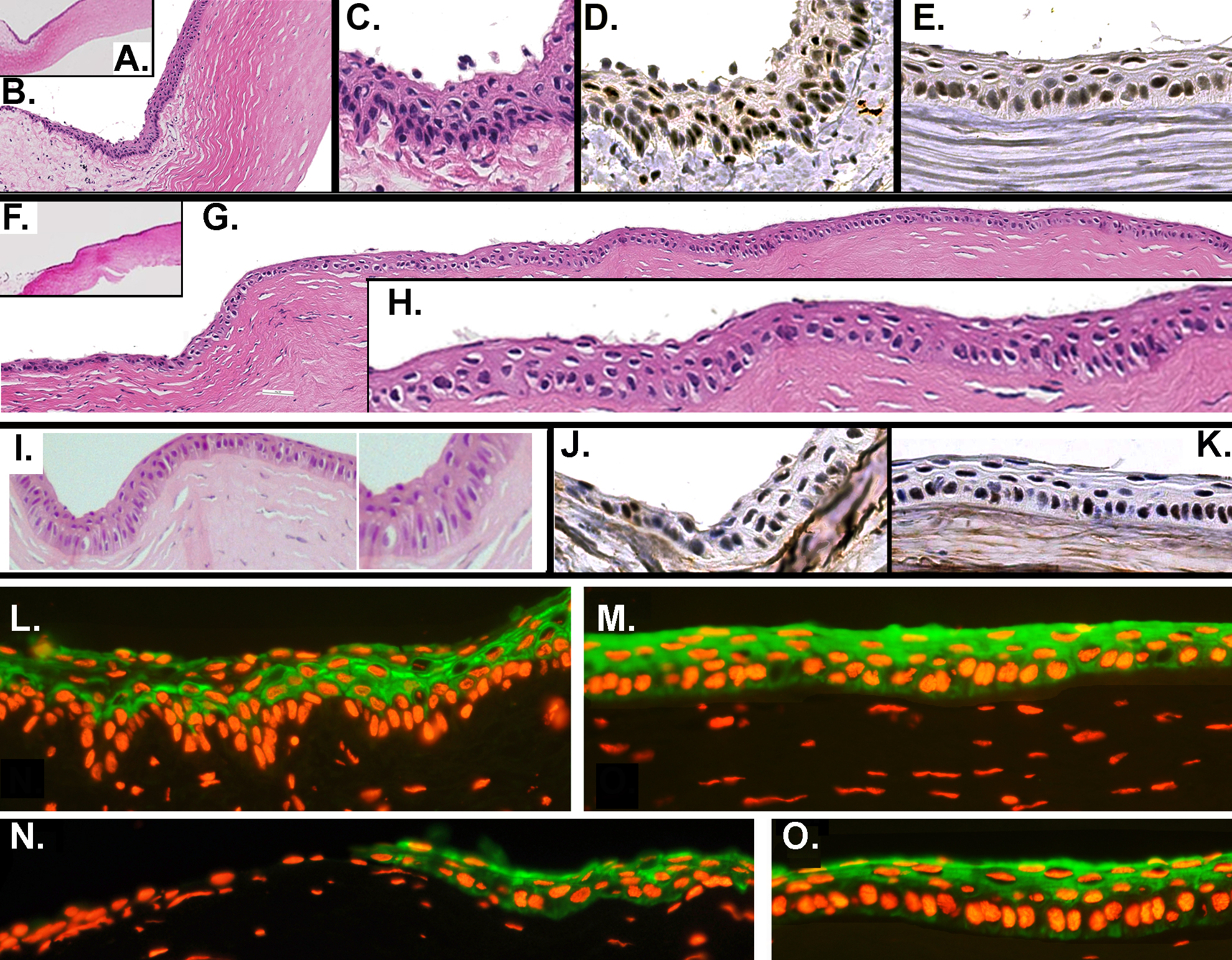
 Figure 5 of
Selver, Mol Vis 2016; 22:138-149.
Figure 5 of
Selver, Mol Vis 2016; 22:138-149.