Figure 4. Photographs of corneas 6 months after excision surgery and grafting of explant outgrowths. A: Human amniotic membrane (hAM) alone. B–D: hAM with outgrowths from the contralateral and donor eye explants. B: Grade 0 neovascularization (neo) and opacity (opc). C: Neo, 1; opc, 0. D: Neo, 2; opc, 2.
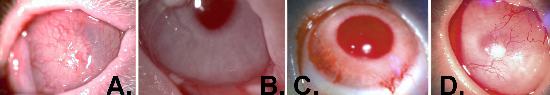
 Figure 4 of
Selver, Mol Vis 2016; 22:138-149.
Figure 4 of
Selver, Mol Vis 2016; 22:138-149.