Figure 3. H&E stainings of the ocular surface immediately after the combined surgical and debridement treatment. A: Composite of micrographs covering the limbal and peripheral zone. The areas subjected to surgical excision and debridement
are indicated. B: Higher magnification of the transition between the surgically excised and debrided areas. C: Area in the central corneal epithelium with remnants of mechanically broken basal cells (arrow in insert). Bar equals 900
μm in A, 300 μm in B and C, and 100 μm in C, insert.
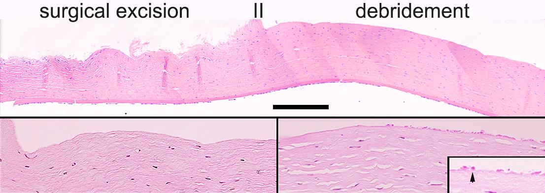
 Figure 3 of
Selver, Mol Vis 2016; 22:138-149.
Figure 3 of
Selver, Mol Vis 2016; 22:138-149.