Figure 4. Angiographic analysis of the effect of Islet-1 siRNA on retinal neovascularization in the mouse. A: Room air–raised mice with normal retinal vessel structure. B: In the murine model of oxygen-induced retinopathy (OIR), neovascular tufts (indicated by the arrow) appear as hyperfluorescence
at the junction between the perfused and non-perfused areas (indicated with a star). C: The murine OIR model with injection of negative control siRNA. Neovascular tufts are apparent at the junction between the
perfused and non-perfused areas. D: The murine OIR model injected with siRNA targeting insulin gene enhancer protein ISL-1 (Islet-1). E: The red area indicates the non-perfusion area. F: The non-perfusion areas were reduced after intravitreal injection of siRNA against Islet-1 compared with that of the murine
model of OIR (★p<0.05).
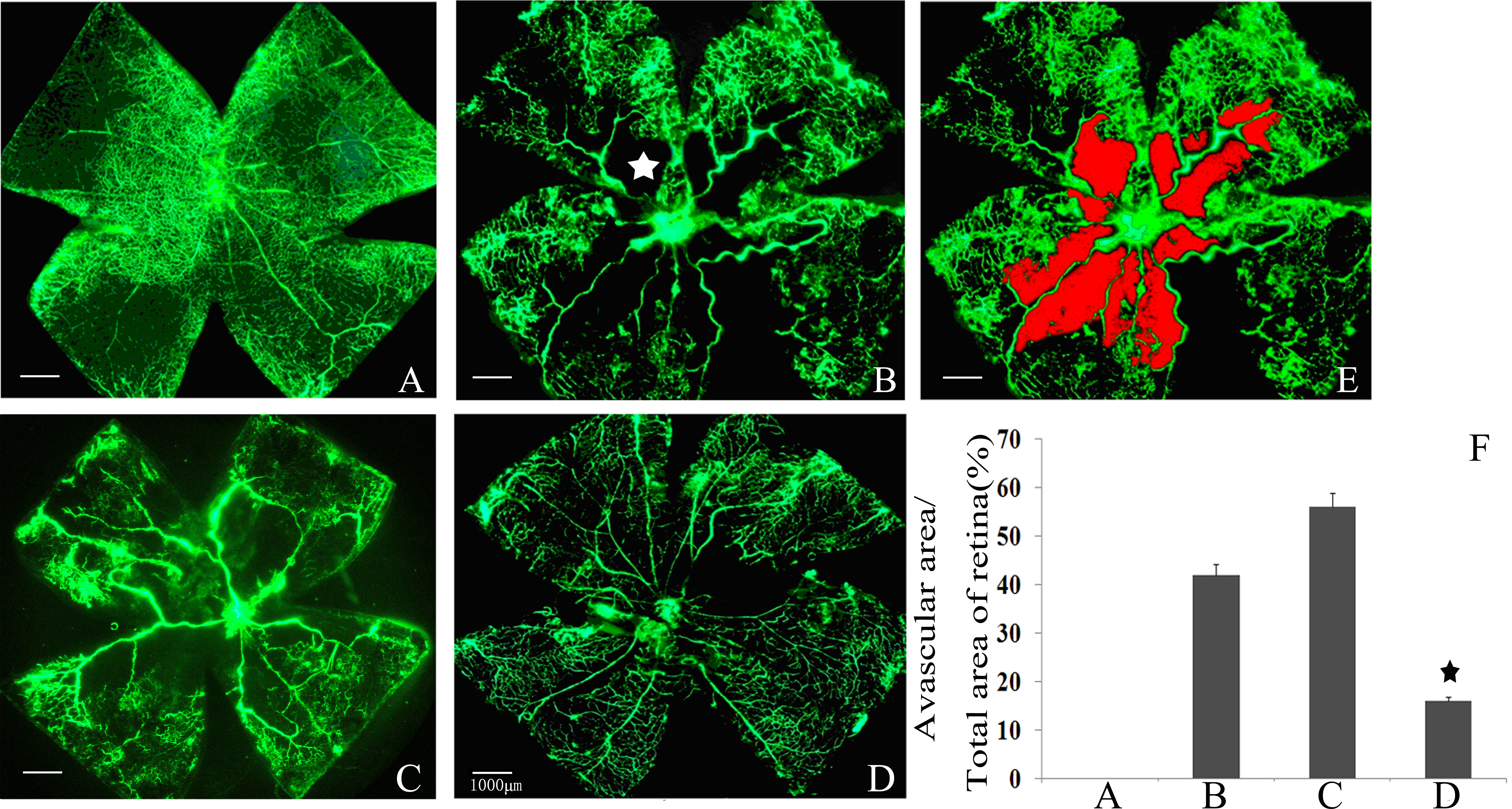
 Figure 4 of
Xiong, Mol Vis 2016; 22:1375-1386.
Figure 4 of
Xiong, Mol Vis 2016; 22:1375-1386.