Figure 8. PKR monoclonal antibody intravitreal injection attenuates CNV. A: Western blot analysis showed that dsRNA-activated protein kinase (PKR), phosphophosphatidylinositol 3-kinase (p-PI3K), phosphoprotein
kinase B (p-Akt), and vascular endothelial growth factor (VEGF) expression in the choroid-RPE-retina complex at day 7 after
laser coagulation. B: Quantification graphs for PKR, p-PI3K, p-Akt, and VEGF to GAPDH. *p<0.01, anti-PKR versus PBS. C: Hyper-fluorescent leakage surrounding the laser spots was weak in the anti-PKR injection mice retinas. D: Representative images of isolectin B4 staining of the RPE-choroid-sclera whole mount from the control and PKR antibody injection
groups 7 days after laser photocoagulation. E: Quantitative measurement of the choroidal neovascularization (CNV) area. *p<0.01, anti-PKR versus PBS.
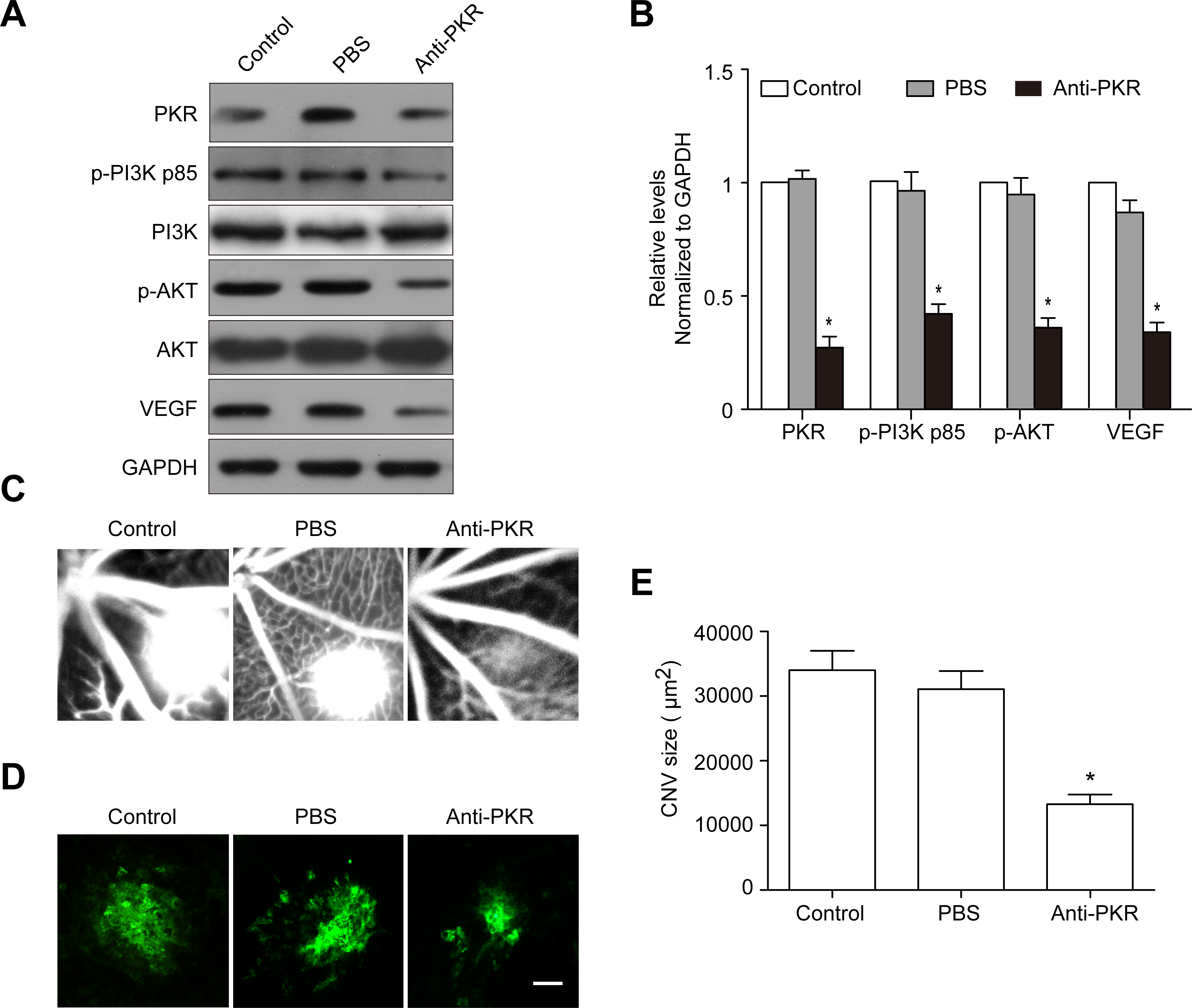
 Figure 8 of
Zhu, Mol Vis 2016; 22:1361-1374.
Figure 8 of
Zhu, Mol Vis 2016; 22:1361-1374.