Figure 4. Localization of GUCY2D LCA1 mutants in mouse retinas. A–D: Confocal retinal membrane
guanylyl cyclase 1 (RetGC1) immunofluorescence images of the GUCY2D-injected human wild-type (A), S248W (B), and R1091x (C) or non-injected Gucy2e−/−Gucy2 knockout (GCdKO) (D) retina sections probed with anti-RetGC1 antibody (red), cone outer segment sheath-staining peanut agglutinin (green), and
TOPRO3 iodide (pseudoblue). The yellow arrowheads point at the cone outer segments surrounding the RetGC1 immunofluorescence.
In all panels, the anti-RetGC1 fluorescence was recorded in the same experiment using identical laser settings. E–G: RetGC1 immunostaining in wild-type (E), S248W (F), and R1091x (G) GUCY2D-expressing retinas shown at lower magnification. Note the faint S248W GUCY2D immunofluorescence in the photoreceptor
cell bodies and axons in the outer nuclear layer (gray arrowheads), along with sparse cone outer segments (yellow arrowheads)
in panel F. Bar length = 50 µm. COS = cone outer segments; ROS = rod outer segments; ONL = outer nuclear layer.
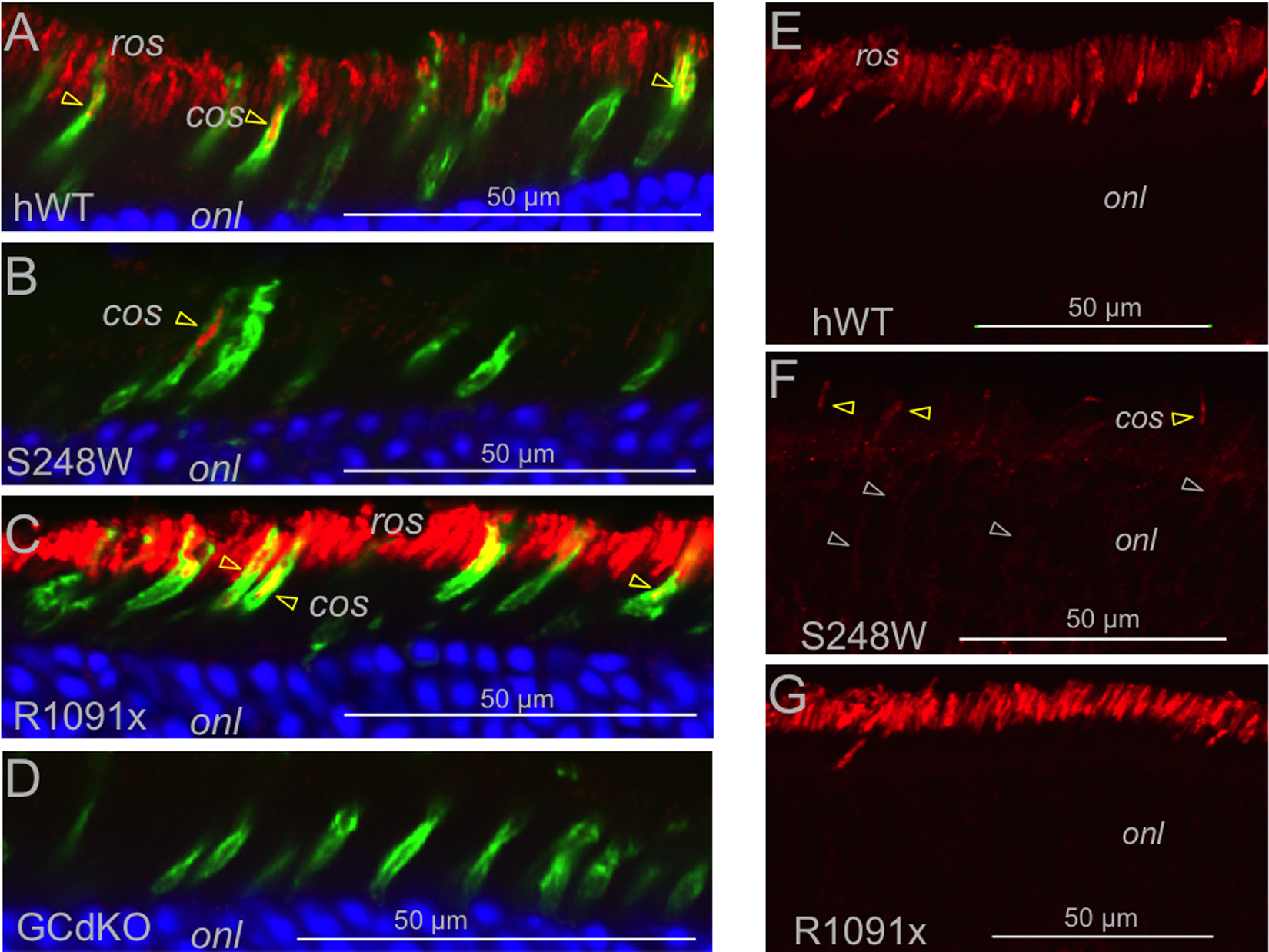
 Figure 4 of
Boye, Mol Vis 2016; 22:1342-1351.
Figure 4 of
Boye, Mol Vis 2016; 22:1342-1351.