Figure 4. Assessment of retinal toxicity 18 h after intravitreal administration of rtPA in rat eyes under excitotoxic conditions. Animals
received either N-methyl-D-aspartate (NMDA; 45 nmol/6 µl) + saline solution or NMDA (45 nmol/6 µl) + recombinant tissue plasminogen
activator (rtPA; 0.27 µg/5 µl). A: Number of annexin V-positive retinal ganglion cells quantified on flatmounted retinas, showing no difference between the
eyes that received NMDA + saline and the eyes that received NMDA + rtPA. B: Number of Brn3a-positive living retinal ganglion cells, quantified on flatmounted retinas, showing no difference between
the eyes that received NMDA + saline and the eyes that received NMDA + rtPA. C: Number of living retinal ganglion cells, quantified on the toluidine blue–stained histologic radial retinal sections, as
the number of cell nuclei visualized at the level of the retinal ganglion cell layer, showing no difference between the eyes
that received NMDA + saline and the eyes that received NMDA + rtPA. D: Toluidine blue–stained retinal radial section illustrating the similar density of retinal ganglion cells in the eyes that
received NMDA + saline (left) and the eyes that received NMDA + rtPA (right). GCL = ganglion cell layer, INL = inner nuclear
layer, ONL = outer nuclear layer. Bar = 20 µm.
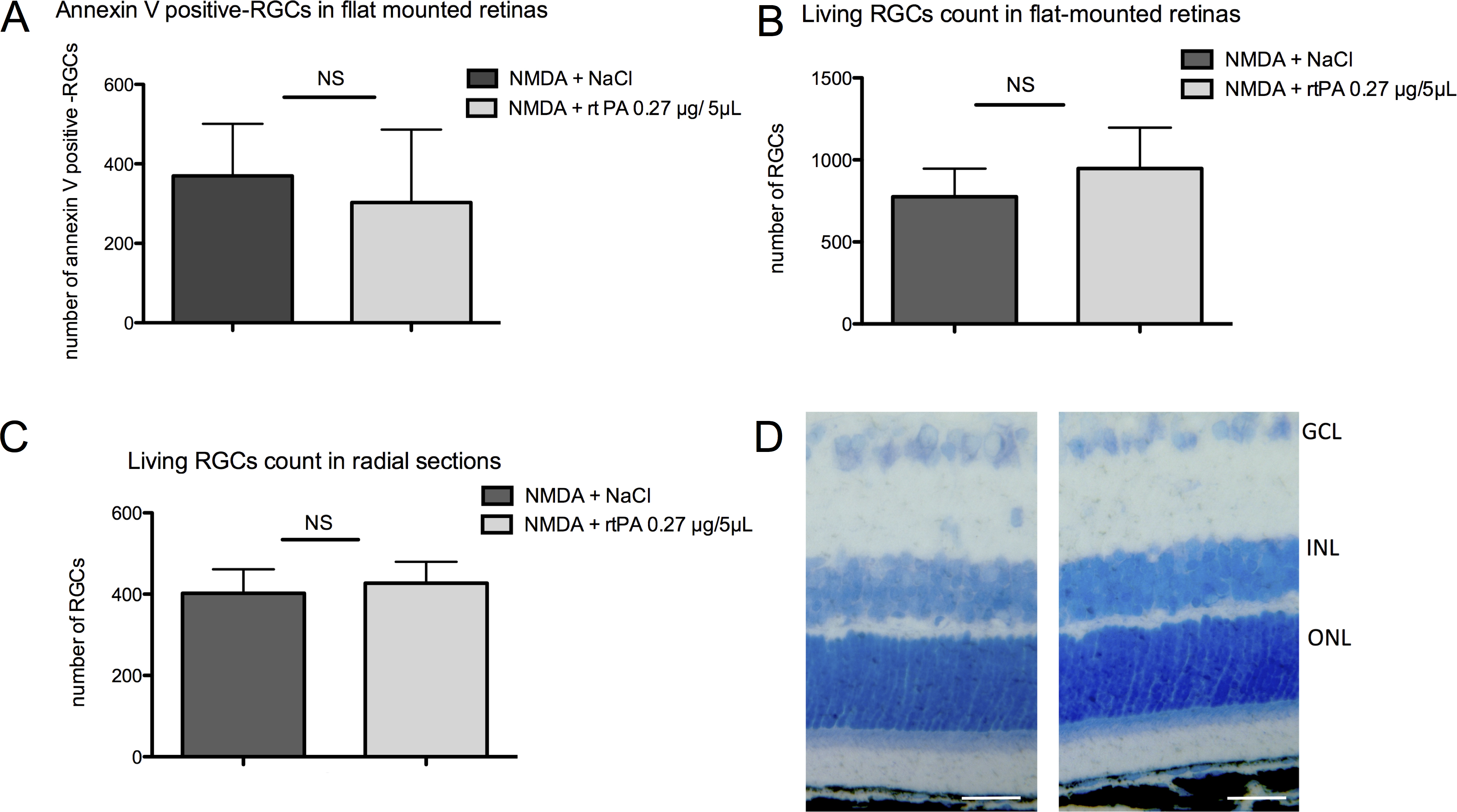
 Figure 4 of
Daruich, Mol Vis 2016; 22:1332-1341.
Figure 4 of
Daruich, Mol Vis 2016; 22:1332-1341.