Figure 7. Localization of HSPA5 and WGA in retinas from human donors with no ocular pathology and age-matched donors with diagnosis
of atrophic AMD. A-F: Localization of HSPA5 and WGA in the retina of a 77-year-old male who had cataract surgery with intraocular lens (IOL) insertion
and no history of AMD. A-C: In the central macular region of a healthy donor retina, the merged image shows the localization of HSPA5 (green) to the
cone IPM in the fovea with lesser labeling around the rod photoreceptors (blue). D-F: Merged image of the peripheral retinal area from the same healthy donor demonstrates scattered labeling of HSPA5 (green)
on the cone and rod photoreceptors (blue). G-L: Localization of HSPA5 and WGA in the retina of a 91-year-old female with dry AMD and bilateral cataracts. G-I: Merged image of the central macular region from a dry AMD donor retina shows negligible HSPA5 (green) staining. J-L: Merged image of the peripheral retinal area from the same donor with dry AMD shows scattered and unstructured HSPA5 (green)
staining. Scale bar = 10 µm. Images are representative of all the donor eyes that were used in the study.
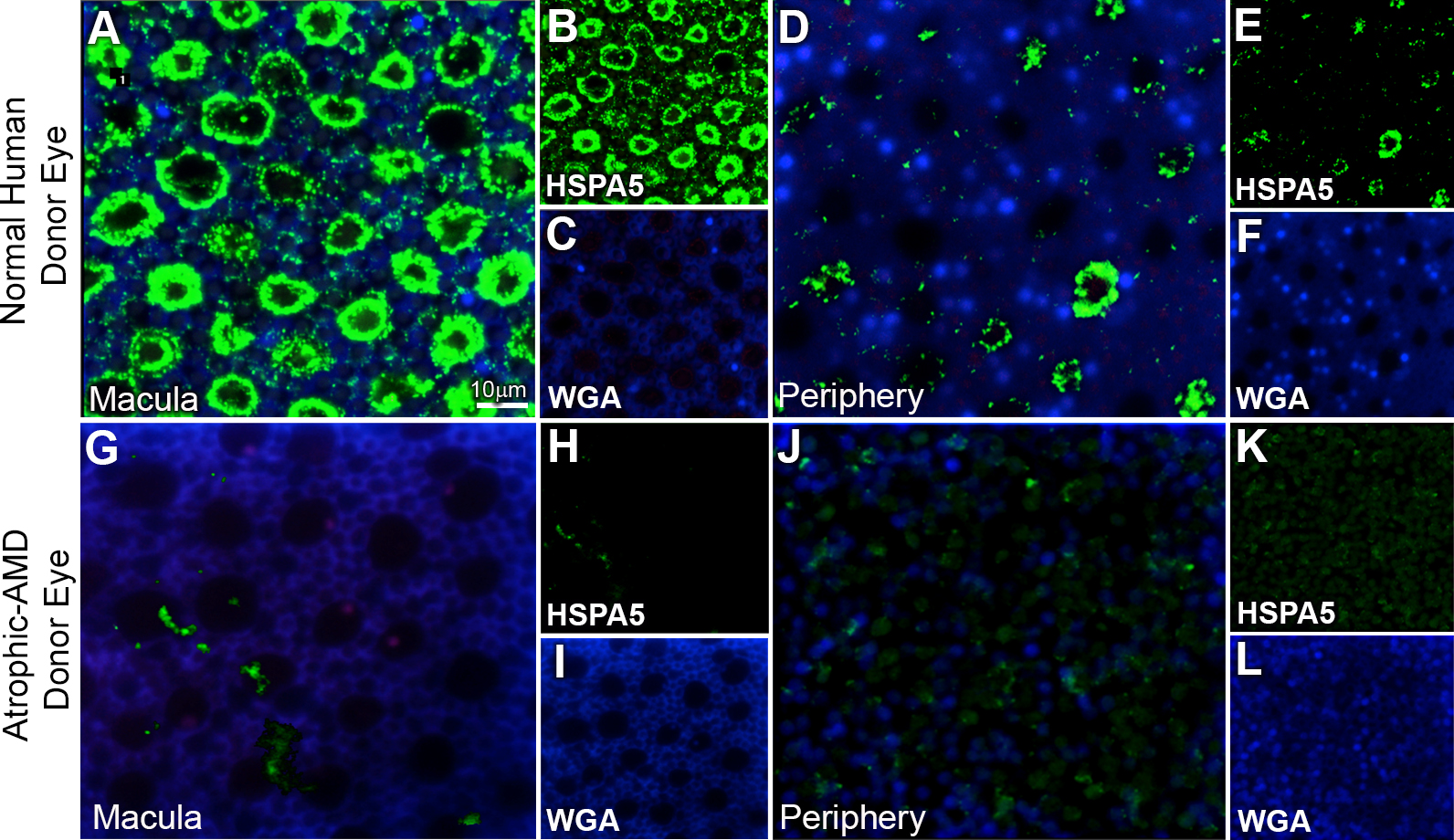
 Figure 7 of
Chintalapudi, Mol Vis 2016; 22:1318-1331.
Figure 7 of
Chintalapudi, Mol Vis 2016; 22:1318-1331.