Figure 6. HSPA5 co-localizes with PNA in flat mounts and cross sections of the human macula with no pathology. A-D: In retinal flat mounts, HSPA5 (green) co-localizes with PNA (red). Punctate yellow indicates co-localization of HSPA5 and
PNA in the IPM of the cone photoreceptors (white arrow). WGA (blue) indicates the IPM of the rods (black arrow). E-H: In retina cross sections, HSPA5 (green) also co-localizes with PNA along the full length of the cones (red, white arrows).
WGA (blue) indicates the IPM of the rods. Scale bar = 10 µm. Electron microscopic immunohistochemical localization of HSPA5
(gold particles) in human retinas (with no ocular pathology). I: HSPA5 is localized to the flocculent material in the IPM surrounding the cone outer segments between adjacent cone and rod
outer segments (box). Scale bar = 2 µm. J: Higher magnification view of HSPA5 localization (black arrow; the boxed area in G is shown magnified in H). COS = cone outer
segment, CIS = cone inner segment, and ROS = rod outer segment. Scale bar = 200 nm. Images are representative of all the donor
eyes that were used in the study.
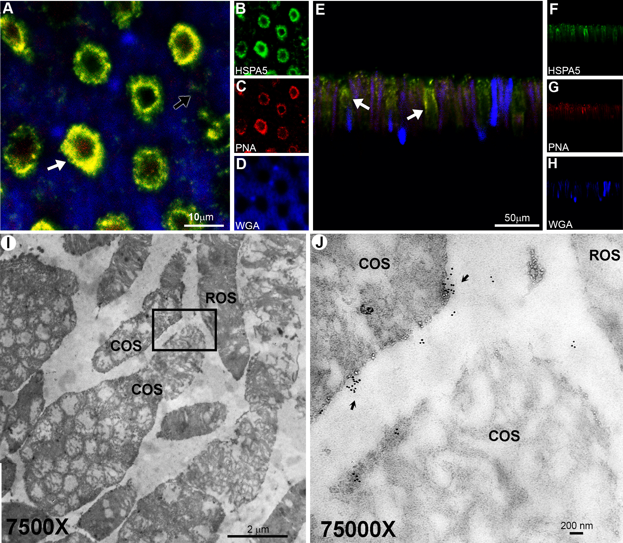
 Figure 6 of
Chintalapudi, Mol Vis 2016; 22:1318-1331.
Figure 6 of
Chintalapudi, Mol Vis 2016; 22:1318-1331.