Figure 5. Immunohistochemical detection of SULF2 in non-pathological mouse and human donor retina cross sections. A-D: In non-permeabilized mouse retina cross sections, SULF2 (red) is observed in the extracellular space around the photoreceptor
inner and outer segments. E-H: In permeabilized mouse retina cross sections, SULF2 (red) is predominantly observed intracellularly in the rod (blue) and
cone (green) photoreceptors, as well as in the cell bodies. I-L: In non-permeabilized non-pathological human retina cross sections, the labeling pattern of SULF2 (red) is identical to that
observed in mice. M-P: In permeabilized human retina cross sections, the SULF2 (red) labeling pattern is also identical to that observed in mice.
OS = outer segments, IN = inner segments, ONL = outer nuclear layer, OPL = outer plexiform layer. Scale bar = 20 µm.
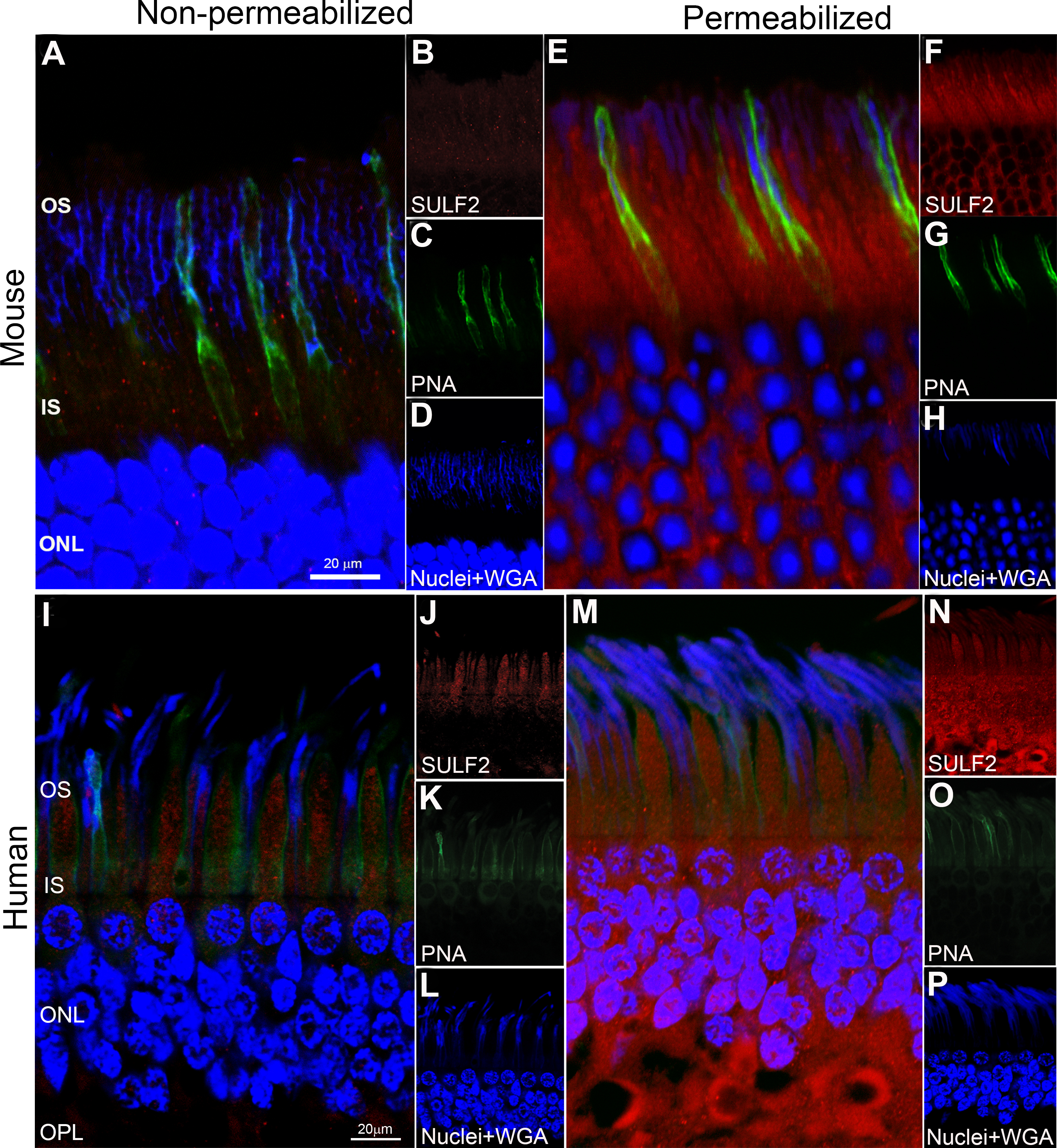
 Figure 5 of
Chintalapudi, Mol Vis 2016; 22:1318-1331.
Figure 5 of
Chintalapudi, Mol Vis 2016; 22:1318-1331.