Figure 1. Immunohistochemical localization of HSPA5 in mice retinas (en face presentation). A-D: HSPA5 (green) co-localizes with PNA (red) in the IPM surrounding the cone photoreceptors. The co-localization of HSPA5 and
PNA is shown in yellow (white arrow). A minimal amount of HSPA5 immunolabeling is associated with WGA around rods (black arrow).
Binding of WGA to the IPM surrounding the rods is shown in blue. Scale bar = 10 µm.
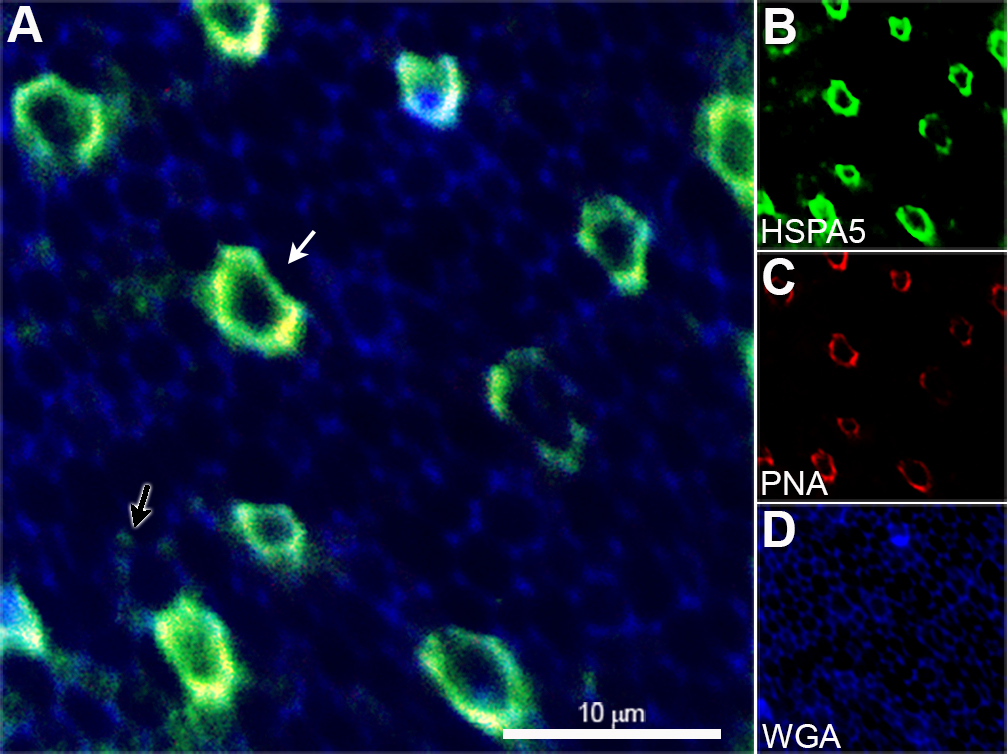
 Figure 1 of
Chintalapudi, Mol Vis 2016; 22:1318-1331.
Figure 1 of
Chintalapudi, Mol Vis 2016; 22:1318-1331.