Figure 2. Sequencing chromatograms of variations in TGFBI identified in this study. A: The c.1663 C>T (p. R555W) variation (heterozygote) in granular corneal dystrophy type 1 (GCD1). B: The c.1663 C>T (p. R555W) variation (homozygote) in GCD1. C: The c.370 T>C (p. R124C) variation (heterozygote) in lattice corneal dystrophy type 1 (LCD1).
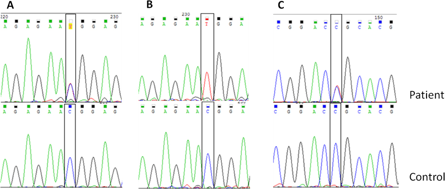
 Figure 2 of
Yaylacioglu Tuncay, Mol Vis 2016; 22:1267-1279.
Figure 2 of
Yaylacioglu Tuncay, Mol Vis 2016; 22:1267-1279.