Figure 4. Analysis of protein expression in human LG and SMG tissue. Proteins from the three donors (D1, D2, and D3) were extracted
using radioimmunoprecipitation assay (RIPA) buffer and processed for sodium dodecyl sulfate–polyacrylamide gel electrophoresis
(SDS–PAGE) or western blotting. A: Protein samples were used for SDS–PAGE and Coomassie staining to investigate the total protein expression and integrity.
B: Western blotting was used to investigate the expression of aquaporin 5, E-cadherin, alpha-smooth muscle actin (α-SMA), and
β-actin in the lacrimal gland (LG) and submandibular gland (SMG) samples.
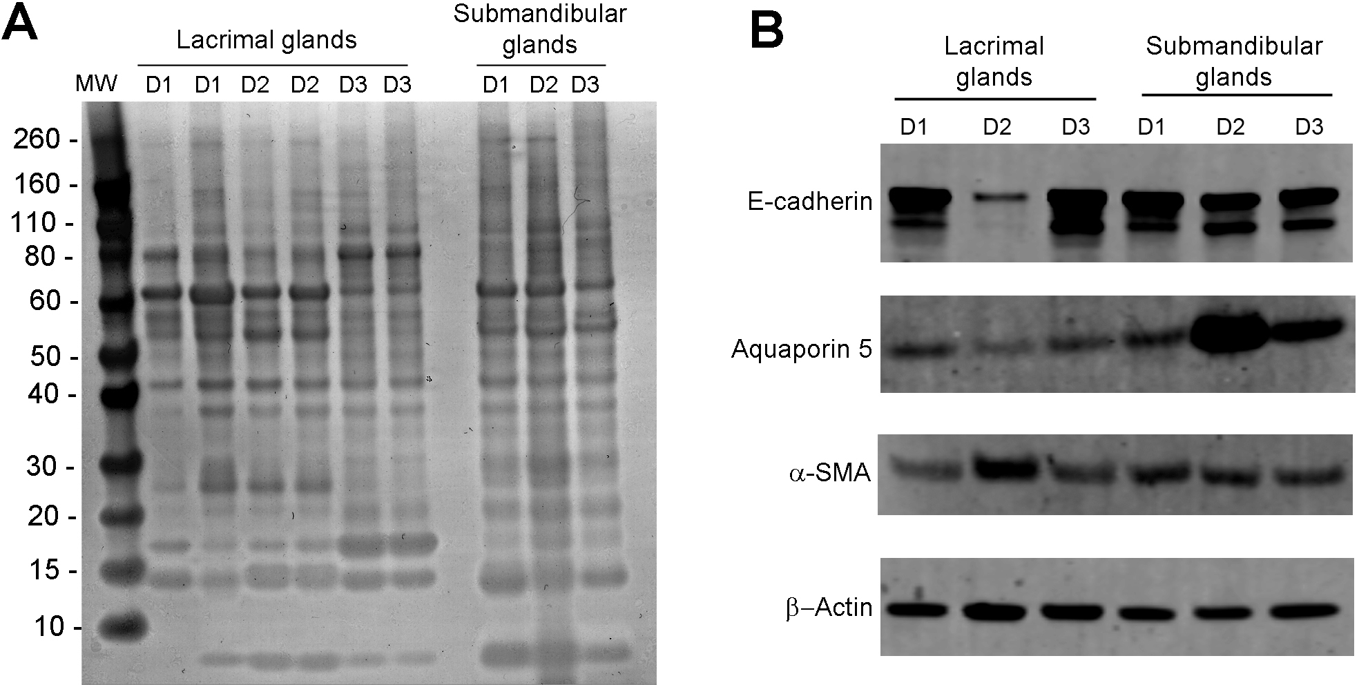
 Figure 4 of
Hawley, Mol Vis 2016; 22:1221-1228.
Figure 4 of
Hawley, Mol Vis 2016; 22:1221-1228.