Figure 3. Immunohistochemical analysis of common LG and SMG cellular proteins. Tissue was fixed and processed for paraffin embedding.
The lacrimal gland (LG) (A, C, E) and submandibular gland (SMG) (B, D, F) sections were stained with antibodies against E-cadherin, aquaporin 5, and alpha-smooth muscle actin (α-SMA) and then counterstained
with 4',6-diamidino-2-phenylindole (DAPI) to stain the nuclei. Images are from a single representative donor. Scale bar =
25 µm. Magnification = 400X.
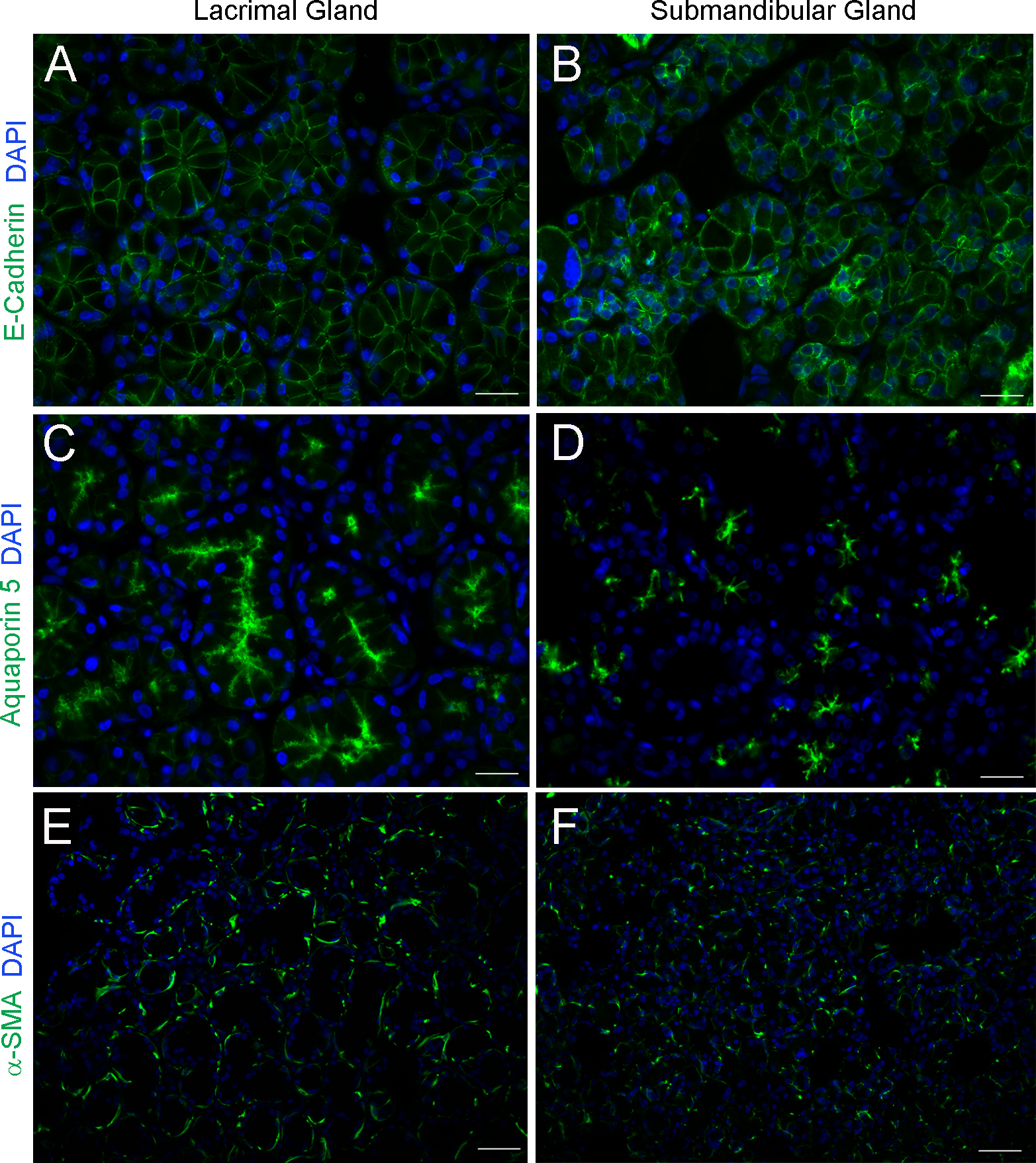
 Figure 3 of
Hawley, Mol Vis 2016; 22:1221-1228.
Figure 3 of
Hawley, Mol Vis 2016; 22:1221-1228.