Figure 2. SCF/cKit induces phosphorylation of Src in human retinal vascular endothelial cells. A–B: Representative western blotting images and densitometric analysis of stem cell factor (SCF)-induced phosphorylation of cKit
and Src are illustrated. In A, human retinal microvascular endothelial cells (HRMECs) treated with imatinib mesylate (10 μM) or untreated cells were stimulated
with rh SCF (50 ng/mL) or PBS for 30 min. Relative intensities of p-cKit and p-Y416 Src were determined by normalization with
band intensity values for total cKit or Src. In B, HRMECs treated with SU6656 (1 μM) or untreated cells were stimulated with rh SCF (50 ng/ml) or PBS for 30 min. Relative
intensities of p-Y416 Src were determined by normalization with band intensity values for total Src (*p<0.05, #p>0.05 versus PBS, n = 3 in A and B). C: Representative immunofluorescence images illustrate the localization of p-Y416 Src. The arrowheads indicate the localization
of p-Y416 Src at the endothelial junctions. HRMECs were pretreated with imatinib mesylate (10 μM) or SU6656 (1 μM) for 30
min before a stimulation with rh SCF (50 ng/ml) or PBS. Cells were stained with immunoglobulin G (IgG) against p-Y416 Src
(green) and VE-cadherin (red). Scale bar = 25 μm.
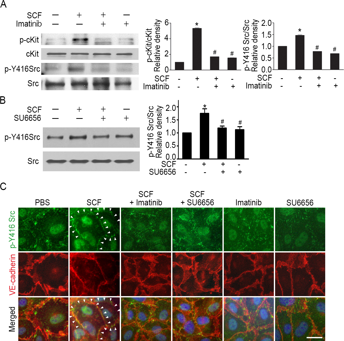
 Figure 2 of
Im, Mol Vis 2016; 22:1213-1220.
Figure 2 of
Im, Mol Vis 2016; 22:1213-1220.