Figure 2. The proliferation of hRECs with endogenous overexpressed TTR. A: TTR was overexpressed in human retinal microvascular endothelial cells (hRECs) with the pCDNA3.1 plasmid in natural medium,
and the expression levels were determined with enzyme-linked immunosorbent assay (ELISA). TTR content in the overexpressed
cells (TTR OE) was always approximately threefold or more compared to that in the control (*:p < 0.001, F = 113.347 (24 h);
p < 0.001, F = 55.084 (48 h); p < 0.001, F = 78.254 (72 h)). B: After the hRECs were cultured with overexpressed TTR for 72 h, only in the hyperglycemia (HG) media, including HG+TTR OE
and HG+Hypoxia+TTR OE, the proliferation of hRECs was significantly inhibited (**: p = 0.003, F = 21.997; ##: p < 0.001, F
= 105.887). In the natural medium (LG) media, the growth of hRECs was slightly enhanced with TTR (*: p = 0.002, F = 27.308;
#: p = 0.002, F = 22.291). (n=3, mean±SD was calculated).
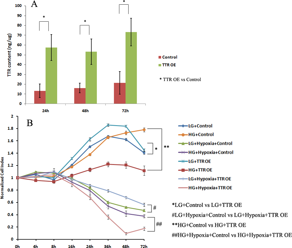
 Figure 2 of
Shao, Mol Vis 2016; 22:1188-1197.
Figure 2 of
Shao, Mol Vis 2016; 22:1188-1197.