Figure 2. Nanoceria do not alter the amount or distribution of phototransduction proteins and caspase 3 level. A: Immunohistochemistry using paraffin sections at PI7 h showed that rhodopsin (red) and M-opsin (green) are properly localized
in the outer segments of the retinas. n = 3–6 eyes per group. Scale bar = 100 µm. B: Western blots were performed at PI7 h to assess the protein levels of rhodopsin, M-opsin and S-opsin, and caspase 3. Densitometric
analysis of the bands is shown as the mean ± standard error of the mean (SEM), and there are no statistically significant
differences among the groups. n = 3–6 eyes per group.
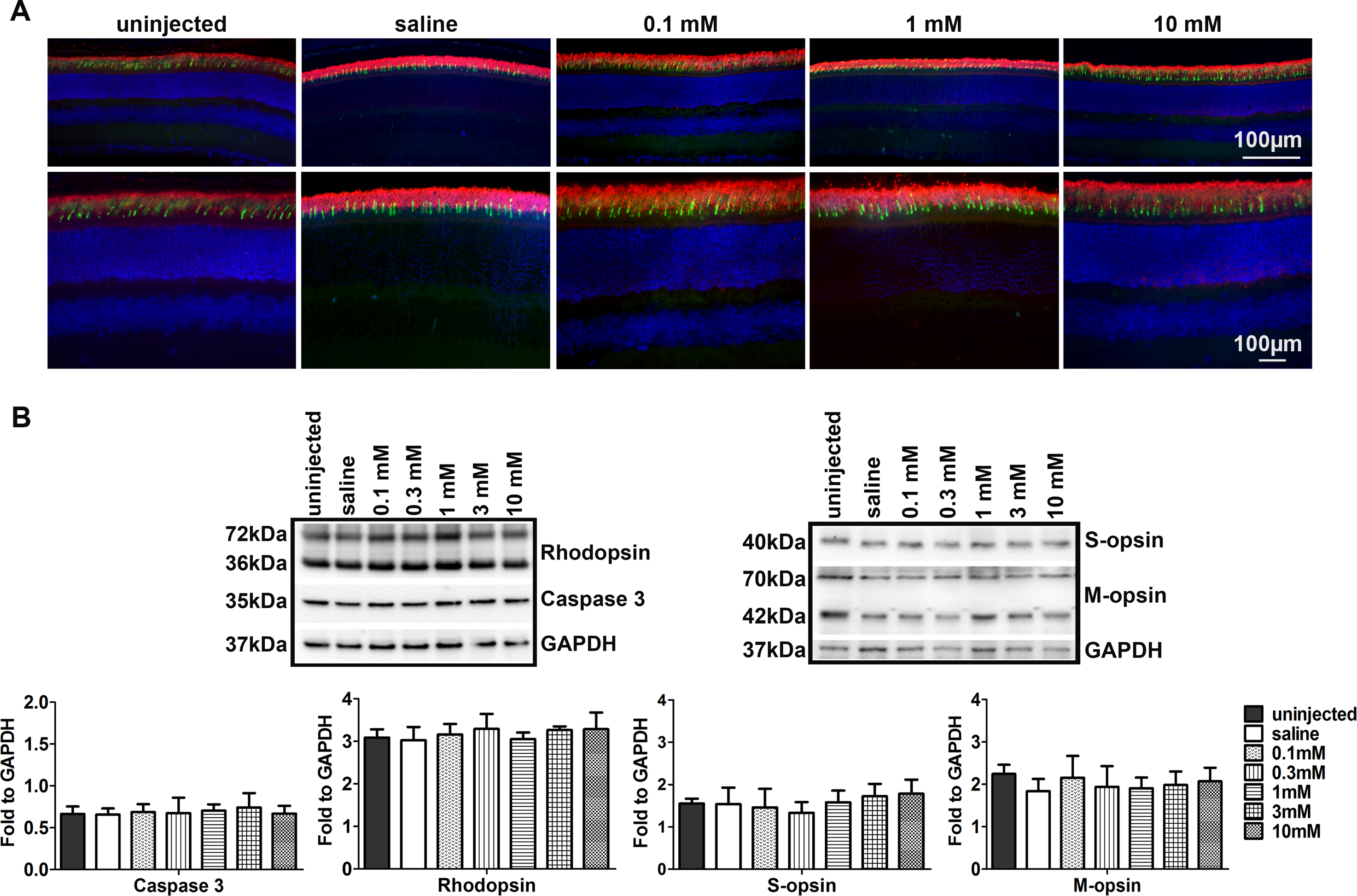
 Figure 2 of
Cai, Mol Vis 2016; 22:1176-1187.
Figure 2 of
Cai, Mol Vis 2016; 22:1176-1187.