Figure 1. Nanoceria do not cause changes in retinal structure or morphology. A: Microscopic images were taken at 0.96 mm from the ONH from the superior side of the retinas using hematoxylin and eosin
(H&E)-stained sections and are representative of three to eight eyes per group. B: Morphometric analysis of the ONL thickness across the entire retinas of each group. Each slide was measured at five points
superiorly and inferiorly under 60X, and the average of the same point from three to eight eyes per group is shown. C: Quantitative histology of the average of 30–80 measurements per group demonstrated that no significant differences occurred
among the groups. RPE, retinal pigment epithelium; OS, outer segment; ONL, outer nuclear layer; INL, inner nuclear layer;
GCL, ganglion cell layer. Scale bar = 100 µm.
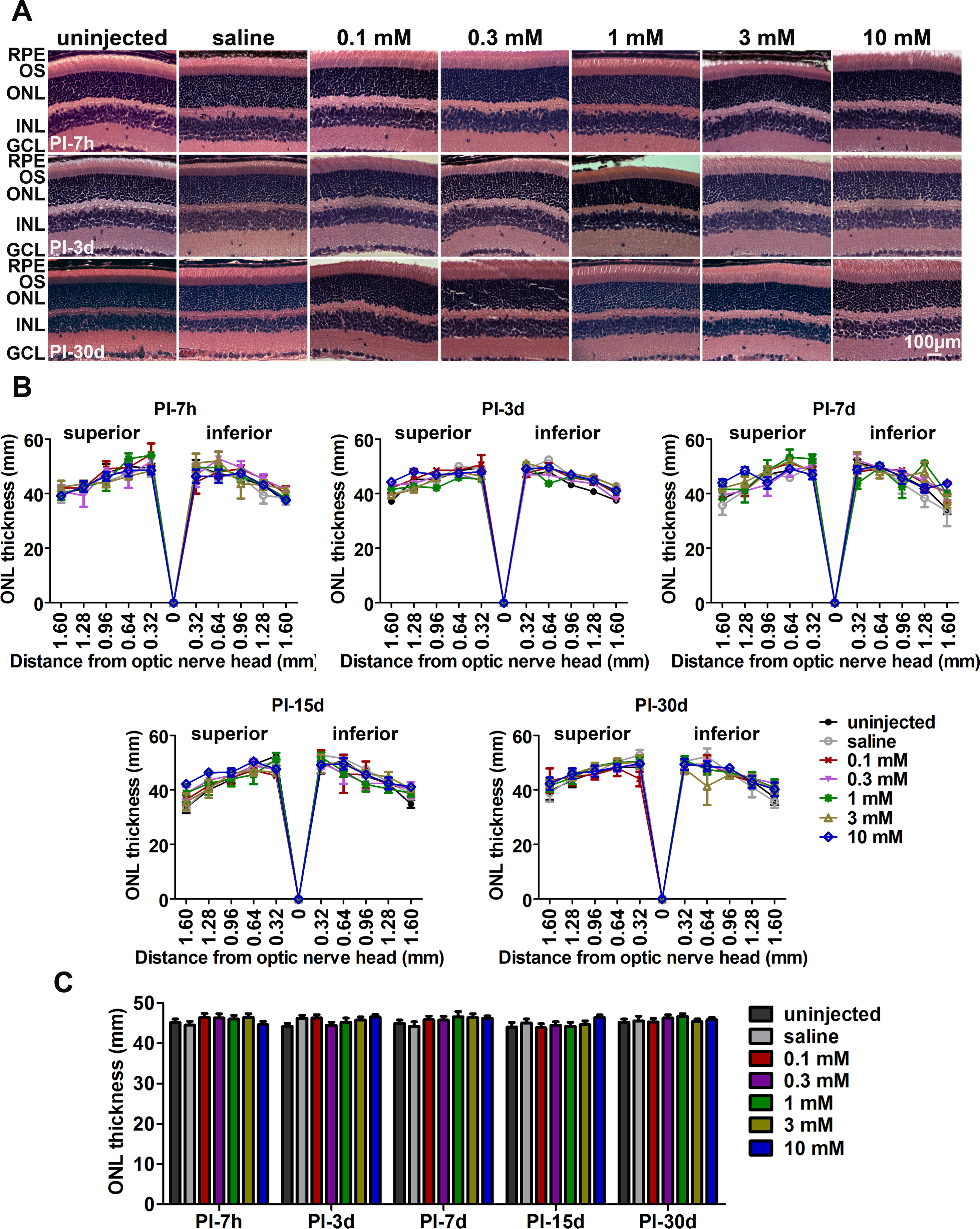
 Figure 1 of
Cai, Mol Vis 2016; 22:1176-1187.
Figure 1 of
Cai, Mol Vis 2016; 22:1176-1187.