Figure 3. TNF-α induces ROS generation in human RPE cells by activating NADPH oxidase. A: Reactive oxygen species (ROS) generation measured with 2',7'-dichlorofluorescein diacetate (DCFDA) fluorescence in human
RPE cells treated with recombinant human tumor necrosis factor alpha (TNF-α) at various concentrations for 30 min (***p<0.001
versus PBS; †††p<0.001 versus TNF-α at 10 ng/ml; ‡‡p<0.01 versus TNF-α at 30 ng/ml). B: ROS generation measured with DCFDA fluorescence in human RPE cells pretreated with apocynin (Apo, 100 µM) and VAS 2870 (VAS,
20 µM) for 30 min before incubation with TNF-α (20 ng/ml) for an additional 30 min (***p<0.001 versus vehicle; †††p<0.001 versus TNF-α). C: Western blot of NOX4. D: Representative gel images of coimmunoprecipitation of NOX4 and p22phox in RPE cells treated with TNF-α or vascular endothelial
growth factor (VEGF) overnight (*p<0.05 versus PBS). E: Quantification of densitometry in RPE cells treated with TNF-α or VEGF overnight (*p<0.05 versus PBS). F: ROS generation measured with DCFDA fluorescence in human RPE cells) with p22phox knockdown by siRNA transfection and incubated
with TNF-α (20 ng/ml) for 30 min (*p<0.05 versus PBS of ConsiRNA; †††p<0.001 versus TNF-α of ConsiRNA).
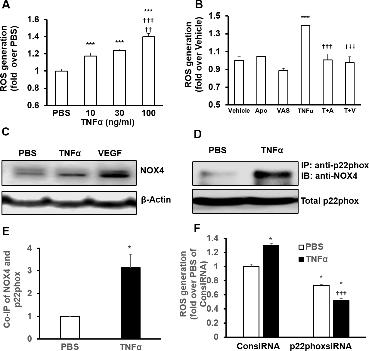
 Figure 3 of
Wang, Mol Vis 2016; 22:116-128.
Figure 3 of
Wang, Mol Vis 2016; 22:116-128.