Figure 1. Effect of proinflammatory cytokines on cultured ARPE-19 cells exhibiting RPE characteristics. The cells were treated with
interferon gamma (IFN-γ; 10 u/ml), tumor necrosis factor alpha (TNF-α; 1 ng/ml), and interleukin-1 beta (IL-1β; 1 ng/ml) for
20 h in the absence of serum and their gene expression analyzed with real-time PCR. A: Control cells exhibited typical epithelial morphology when examined with phase contrast microscopy. Cells treated with cytokines
appeared distorted with thickened cell junctions. Magnification: 100X. B: The cells responded to the proinflammatory cytokines by highly increasing the expression of cytokines and chemokines. Gene
expression in the treated and control cells was analyzed with real-time PCR. The fold increases shown are statistically significant
(p<0.05 compared to control, n = 4). C: The expression of the RPE characteristic genes when analyzed with real-time PCR decreased substantially in the differentiated
ARPE-19 cells exposed to the proinflammatory cytokines. *p<0.5 when compared to control, n = 4.
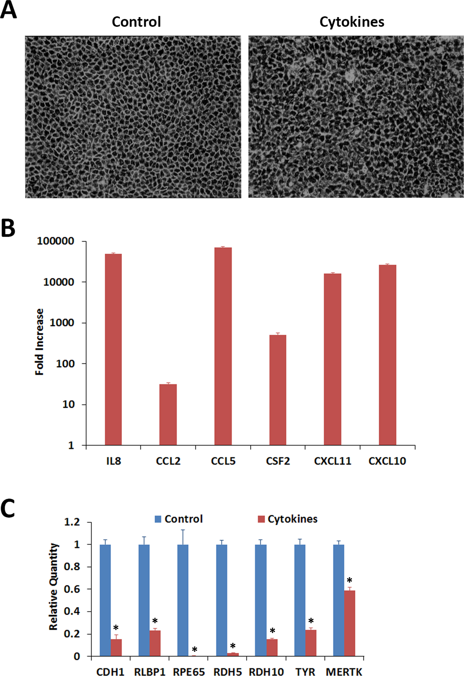
 Figure 1 of
Kutty, Mol Vis 2016; 22:1156-1168.
Figure 1 of
Kutty, Mol Vis 2016; 22:1156-1168.