Figure 6. High magnification images of citrullinated GFAP filaments in the injured retina. Tissue sections of mouse eyes injured for
either 3 h or 3 days were immunostained for GFAP (A, E, red), citrullinated proteins (B, F, F95 antibody, green), and nuclei (C,G, merged images, DAPI-blue). Sections were analyzed on a Zeiss LSM 780 confocal microscope at 63X magnification to identify
GFAP filaments co-localized with citrullinated species. Colocalized filaments in 3 h (C, D, asterisk) and 3 day samples (G, H, asterisk) were visually identifiable in the inner plexiform layer (IPL). Orthogonal planes were analyzed (D, H) to identify points of co-localization (asterisks, arrowheads). Invasion of filaments into the vitreal space past the ganglion
cell layer (GCL) was identified in 3 h injured samples (C, bracket). (Scale bar = 40 μm).
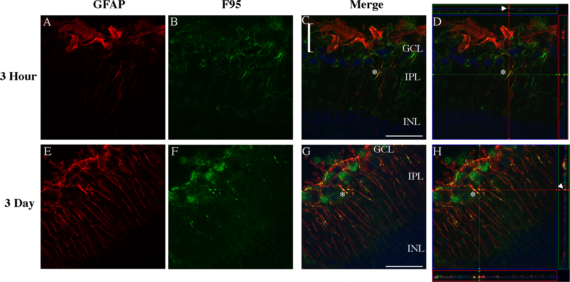
 Figure 6 of
Wizeman, Mol Vis 2016; 22:1137-1155.
Figure 6 of
Wizeman, Mol Vis 2016; 22:1137-1155.