Figure 5. Citrullination along vimentin filaments after ocular injury. Cryosections from uninjured and injured eyes were stained using
antibodies against vimentin (red) and citrullinated proteins (F95; green). Tissue sections were examined under confocal microscope
at 20X magnification from uninjured eyes (A-C) and injured eyes at 1 h (D-F), 3 h (G-I), 1 day (J-L) and 3 days (M-O) postinjury. Extensions of vimentin staining beyond nuclei of GCL are marked by arrow (I, O). Representative regions showing the overlap of vimentin and F95 antibodies are marked by asterisks (F, I, O). Interspersed regions of vimentin and F95 reactivity are outlined in brackets (F, I, O). Diffuse staining of F95 is marked by an arrowhead (O). GCL, ganglion cell layer; IPL, inner plexiform layer; INL, inner nuclear layer; OPL, outer plexiform layer; ONL, outer
nuclear layer. (Scale bar = 100 μm).
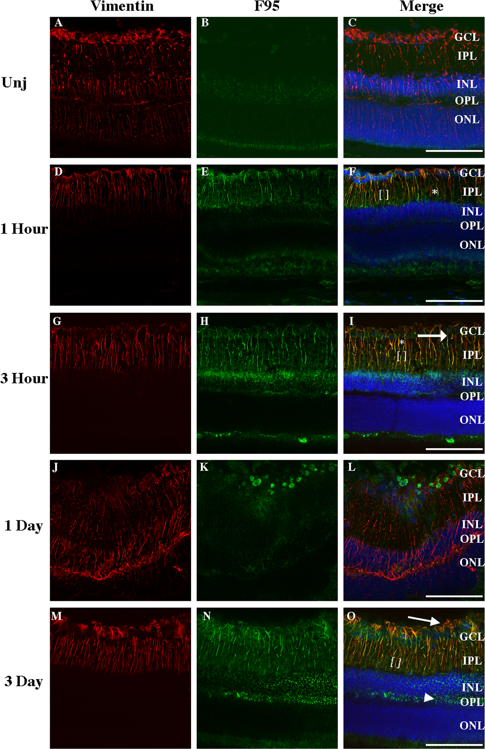
 Figure 5 of
Wizeman, Mol Vis 2016; 22:1137-1155.
Figure 5 of
Wizeman, Mol Vis 2016; 22:1137-1155.