Figure 4. Citrullination of GFAP filaments after injury. Cryosections from uninjured and injured eyes were stained with GFAP (red) and
F95 (green) and nuclei stained with DAPI (blue). Sections were examined under confocal microscope at 20X magnification in
an uninjured state (A-C) and at 1 h (D-F), 3 h (G-I), 1 day (J-L) and 3 days (M-O) postinjury. Extensions of GFAP staining beyond nuclei of the GCL are marked by an arrow (F). Co-localization of GFAP and F95 antibodies are marked by asterisks (F, I, L, O). Interspersed regions of GFAP and F95 reactivity is outlined in brackets (I, O). GFAP-negative, F95-positive filaments are marked by arrowhead (I). GCL, ganglion cell layer; IPL, inner plexiform layer; INL, inner nuclear layer; OPL, outer plexiform layer; ONL, outer
nuclear layer. (Scale bar = 100 μm).
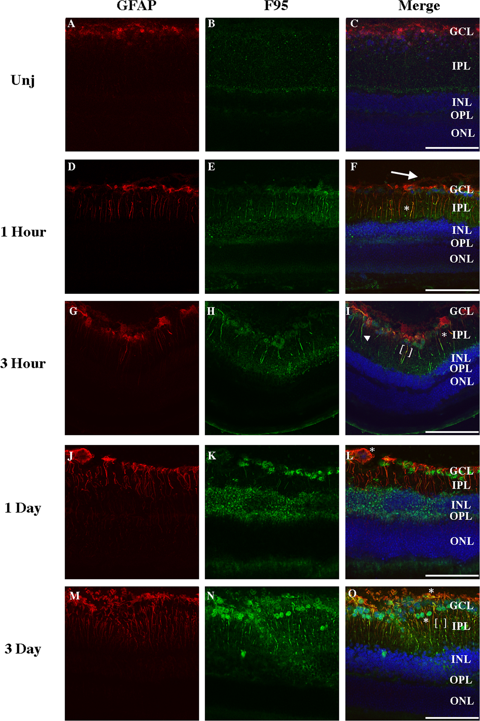
 Figure 4 of
Wizeman, Mol Vis 2016; 22:1137-1155.
Figure 4 of
Wizeman, Mol Vis 2016; 22:1137-1155.