Figure 3. Detection of citrullinated GFAP in uninjured and injured retinas. The figure shows two-dimensional (2D) IEF-PAGE separation
of soluble proteins from uninjured and 7 days postinjury retinochoroidal tissues immunoprecipitated for citrullinated proteins
(F95 antibody) and western blotted. A, B: CTGF-1221 antibody was employed first for antigen detection. C, D: F95 antibody was subsequently employed for antigen detection after removal of CTGF-1221 antibody probe. Blots of the injured
retinas probed with the anti-citrullinated GFAP antibody (B) showed lower relative abundance of ~50 kDa protein species compared to 75 kDa species when re-probed with F95 antibody (D).
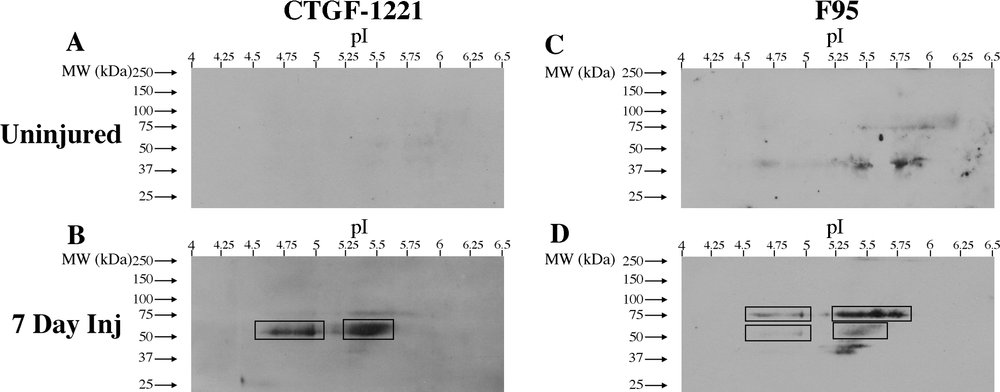
 Figure 3 of
Wizeman, Mol Vis 2016; 22:1137-1155.
Figure 3 of
Wizeman, Mol Vis 2016; 22:1137-1155.