Figure 1. Detection of soluble citrullinated proteins after ocular alkali injury. Western blot analysis of soluble protein extracts
from retinochoroidal tissues of uninjured eyes (lane 1) and mouse eyes injured in vivo for 1, 3, and 7 days. A: Blots were probed for glial fibrillary acidic protein (GFAP). B: Blots were probed for vimentin. C: Blots were probed for citrullinated proteins (F95 antibody). All blots were probed for glyceraldehyde 3-phosphate dehydrogenase
(GAPDH) as a loading control. The major 50-kDa band (C, asterisk), and 75-kDa band (C, arrowhead), along with minor high-molecular-weight-bands over 150- to 250-kDa bands (bracket) were identified at all stages
postinjury when examined for citrullination. GFAP antibody reactive species were detected between 37 kDa and 50 kDa (A). Uninjured and injured eyes exhibit vimentin reactivity at isoforms ranging from 37 to 55 kDa (B). D, E: Bar graphs represent quantification of the 75- and 50-kDa citrullinated species. Each sample contained tissue extracts pooled
from three retinas from which soluble protein was extracted as described in Methods. Experiments were repeated three times
(n =3). Data are expressed as the mean ± standard deviation (SD). P values less than 0.01 (hashtag) as well as less than 0.05
(asterisk) were considered significant as determined by t test.
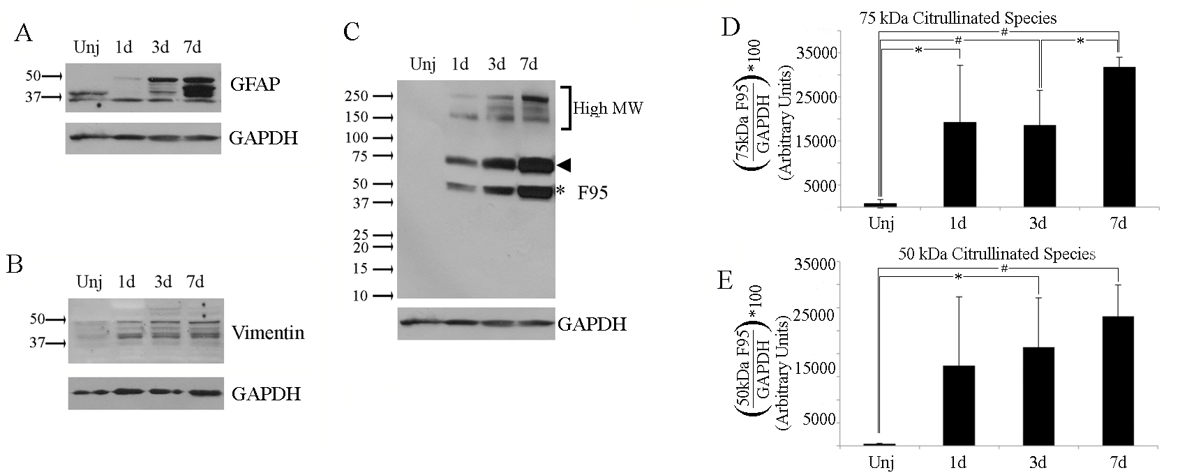
 Figure 1 of
Wizeman, Mol Vis 2016; 22:1137-1155.
Figure 1 of
Wizeman, Mol Vis 2016; 22:1137-1155.