Figure 9. p27 expression in primary cultures of RPE cells. A: Double immunofluorescence for myosin VIIa (MyoVIIa) and α-smooth muscle actin (α-SMA) showing the epithelial–mesenchymal
transition (EMT) of RPE cells in culture. Arrows indicate α-SMA-positive RPE cells while arrowheads denote α-SMA-negative
RPE cells. B: Double immunofluorescence for p27 and ZO-1. p27 is detected in the nucleus (arrowhead) and the cytoplasm (arrows) at day
1 while nuclear and cytoplasmic p27 is lost in many RPE cells by day 3 (double arrows). Hoechst, nuclear staining with Hoechst
33258. Scale bars = 20 μm.
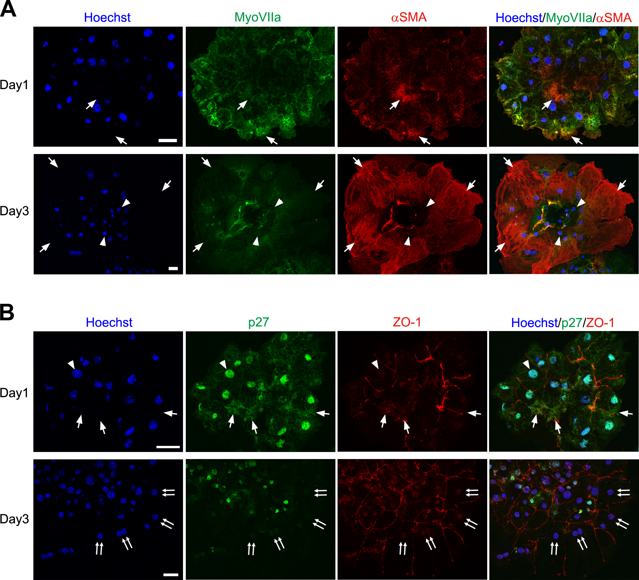
 Figure 9 of
ul Quraish, Mol Vis 2016; 22:1103-1121.
Figure 9 of
ul Quraish, Mol Vis 2016; 22:1103-1121.